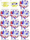
Structural allele-specific patterns adopted by epitopes in the MHC-I cleft and reconstruction of MHC:peptide complexes to cross-reactivity assessment
- PMID: 20442757
- PMCID: PMC2860844
- DOI: 10.1371/journal.pone.0010353
Structural allele-specific patterns adopted by epitopes in the MHC-I cleft and reconstruction of MHC:peptide complexes to cross-reactivity assessment
Abstract
The immune system is engaged in a constant antigenic surveillance through the Major Histocompatibility Complex (MHC) class I antigen presentation pathway. This is an efficient mechanism for detection of intracellular infections, especially viral ones. In this work we describe conformational patterns shared by epitopes presented by a given MHC allele and use these features to develop a docking approach that simulates the peptide loading into the MHC cleft. Our strategy, to construct in silico MHC:peptide complexes, was successfully tested by reproducing four different crystal structures of MHC-I molecules available at the Protein Data Bank (PDB). An in silico study of cross-reactivity potential was also performed between the wild-type complex HLA-A2-NS31073 and nine MHC:peptide complexes presenting alanine exchange peptides. This indicates that structural similarities among the complexes can give us important clues about cross reactivity. The approach used in this work allows the selection of epitopes with potential to induce cross-reactive immune responses, providing useful tools for studies in autoimmunity and to the development of more comprehensive vaccines.
Conflict of interest statement
Figures


Similar articles
-
DockTope: a Web-based tool for automated pMHC-I modelling.Sci Rep. 2015 Dec 17;5:18413. doi: 10.1038/srep18413. Sci Rep. 2015. PMID: 26674250 Free PMC article.
-
Toward the prediction of class I and II mouse major histocompatibility complex-peptide-binding affinity: in silico bioinformatic step-by-step guide using quantitative structure-activity relationships.Methods Mol Biol. 2007;409:227-45. doi: 10.1007/978-1-60327-118-9_16. Methods Mol Biol. 2007. PMID: 18450004
-
In silico analysis of MHC-I restricted epitopes of Chikungunya virus proteins: Implication in understanding anti-CHIKV CD8(+) T cell response and advancement of epitope based immunotherapy for CHIKV infection.Infect Genet Evol. 2015 Apr;31:118-26. doi: 10.1016/j.meegid.2015.01.017. Epub 2015 Jan 31. Infect Genet Evol. 2015. PMID: 25643869
-
T cell antigen receptor recognition of antigen-presenting molecules.Annu Rev Immunol. 2015;33:169-200. doi: 10.1146/annurev-immunol-032414-112334. Epub 2014 Dec 10. Annu Rev Immunol. 2015. PMID: 25493333 Review.
-
Structural Prediction of Peptide-MHC Binding Modes.Methods Mol Biol. 2022;2405:245-282. doi: 10.1007/978-1-0716-1855-4_13. Methods Mol Biol. 2022. PMID: 35298818 Review.
Cited by
-
Structure-based Methods for Binding Mode and Binding Affinity Prediction for Peptide-MHC Complexes.Curr Top Med Chem. 2018;18(26):2239-2255. doi: 10.2174/1568026619666181224101744. Curr Top Med Chem. 2018. PMID: 30582480 Free PMC article. Review.
-
Hepatitis E Virus (HEV)-Specific T Cell Receptor Cross-Recognition: Implications for Immunotherapy.Front Immunol. 2019 Sep 4;10:2076. doi: 10.3389/fimmu.2019.02076. eCollection 2019. Front Immunol. 2019. PMID: 31552033 Free PMC article.
-
HLA3DB: comprehensive annotation of peptide/HLA complexes enables blind structure prediction of T cell epitopes.Nat Commun. 2023 Oct 10;14(1):6349. doi: 10.1038/s41467-023-42163-z. Nat Commun. 2023. PMID: 37816745 Free PMC article.
-
Machine Learning for Cancer Immunotherapies Based on Epitope Recognition by T Cell Receptors.Front Genet. 2019 Nov 19;10:1141. doi: 10.3389/fgene.2019.01141. eCollection 2019. Front Genet. 2019. PMID: 31798635 Free PMC article. Review.
-
DockTope: a Web-based tool for automated pMHC-I modelling.Sci Rep. 2015 Dec 17;5:18413. doi: 10.1038/srep18413. Sci Rep. 2015. PMID: 26674250 Free PMC article.
References
-
- Yewdell JW, Bennink JR. Immunodominance in major histocompatibility complex class I-restricted T lymphocyte responses. Annu Rev Immunol. 1999;17:51–88. - PubMed
-
- Welsh RM, Selin LK, Szomolanyi-Tsuda E. Immunological memory to viral infections. Annu Rev Immunol. 2004;22:711–743. - PubMed
-
- Wilson DB, Wilson DH, Schroder K, Pinilla C, Blondelle S, et al. Specificity and degeneracy of T cells. Mol Immunol. 2004;40:1047–1055. - PubMed
-
- Welsh RM, Selin LK. No one is naive: the significance of heterologous T-cell immunity. Nat Rev Immunol. 2002;2:417–426. - PubMed
Publication types
MeSH terms
Substances
LinkOut - more resources
Full Text Sources
Research Materials

