

Metastasis-inducing S100A4 and RANTES cooperate in promoting tumor progression in mice
- PMID: 20442771
- PMCID: PMC2860983
- DOI: 10.1371/journal.pone.0010374
Metastasis-inducing S100A4 and RANTES cooperate in promoting tumor progression in mice
Abstract
Background: The tumor microenvironment has been described as a critical milieu determining tumor growth and metastases. A pivotal role of metastasis-inducing S100A4 in the development of tumor stroma has been proven in animal models and verified in human breast cancer biopsies. Expression and release of S100A4 has been shown in various types of stroma composing cells, including fibroblasts and immune cells. However, the events implicated in upstream and downstream pathways regulating the activity of the extracellular S100A4 protein in the tumor milieu remain unsolved.
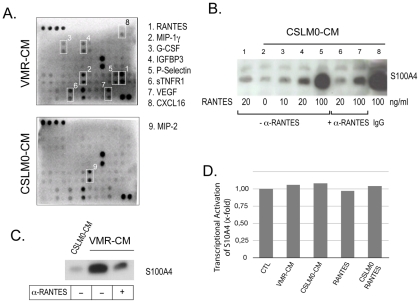
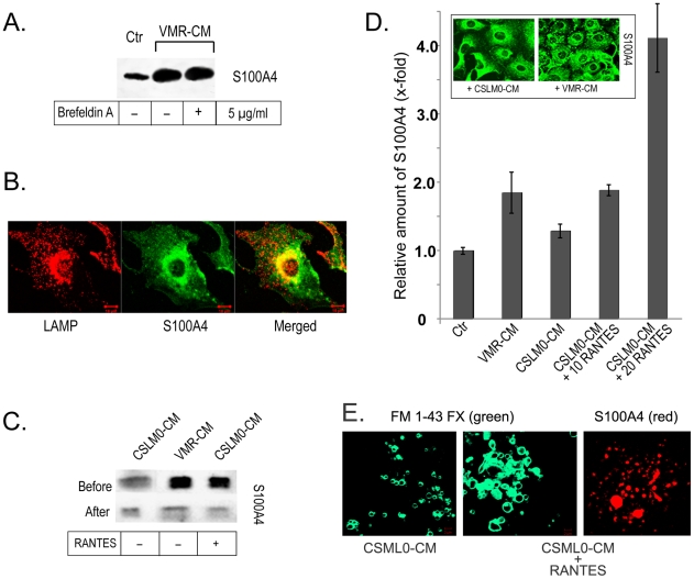
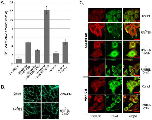
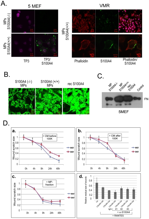
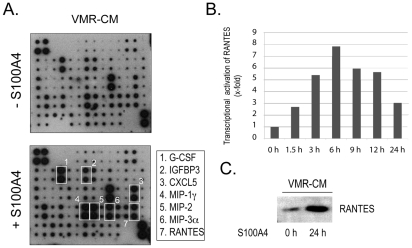
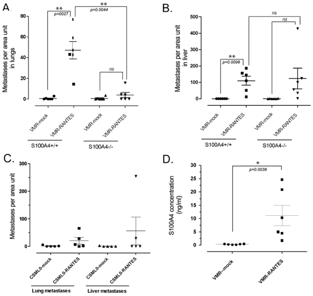
Methodology/principal findings: We studied the interplay between the tumor cell-derived cytokine regulated-upon-activation, normal T-cell expressed and secreted (RANTES; CCL5) and S100A4 which were shown to be critical factors in tumor progression. We found that RANTES stimulates the externalization of S100A4 via microparticle shedding from the plasma membrane of tumor and stroma cells. Conversely, the released S100A4 protein induces the upregulation of fibronectin (FN) in fibroblasts and a number of cytokines, including RANTES in tumor cells as well as stimulates cell motility in a wound healing assay. Importantly, using wild type and S100A4-deficient mouse models, we demonstrated a substantial influence of tumor cell-derived RANTES on S100A4 release into blood circulation which ultimately increases the metastatic burden in mice.
Conclusions/significance: Altogether, the data presented strongly validate the pro-metastatic function of S100A4 in the tumor microenvironment and define how the tumor cell-derived cytokine RANTES acts as a critical regulator of S100A4-dependent tumor cell dissemination. Additionally, for the first time we demonstrated the mechanism of S100A4 release associated with plasma membrane microparticle shedding from various cells types.
Conflict of interest statement
Figures
Similar articles
-
Functional significance of metastasis-inducing S100A4(Mts1) in tumor-stroma interplay.J Biol Chem. 2004 Jun 4;279(23):24498-504. doi: 10.1074/jbc.M400441200. Epub 2004 Mar 26. J Biol Chem. 2004. PMID: 15047714
-
A link between inflammation and metastasis: serum amyloid A1 and A3 induce metastasis, and are targets of metastasis-inducing S100A4.Oncogene. 2015 Jan 22;34(4):424-35. doi: 10.1038/onc.2013.568. Epub 2014 Jan 27. Oncogene. 2015. PMID: 24469032
-
S100A4-neutralizing antibody suppresses spontaneous tumor progression, pre-metastatic niche formation and alters T-cell polarization balance.BMC Cancer. 2015 Feb 12;15:44. doi: 10.1186/s12885-015-1034-2. BMC Cancer. 2015. PMID: 25884510 Free PMC article.
-
Genetically modified mouse models to study the role of metastasis-promoting S100A4(mts1) protein in metastatic mammary cancer.J Dairy Res. 2005;72 Spec No:27-33. doi: 10.1017/s0022029905001093. J Dairy Res. 2005. PMID: 16180718 Review.
-
Metastasis-associated protein S100A4: spotlight on its role in cell migration.Curr Cancer Drug Targets. 2007 May;7(3):217-28. doi: 10.2174/156800907780618329. Curr Cancer Drug Targets. 2007. PMID: 17504119 Review.
Cited by
-
S100 proteins in cancer.Nat Rev Cancer. 2015 Feb;15(2):96-109. doi: 10.1038/nrc3893. Nat Rev Cancer. 2015. PMID: 25614008 Free PMC article. Review.
-
Extracellular S100A4 as a key player in fibrotic diseases.J Cell Mol Med. 2020 Jun;24(11):5973-5983. doi: 10.1111/jcmm.15259. Epub 2020 Apr 19. J Cell Mol Med. 2020. PMID: 32307910 Free PMC article. Review.
-
Leukocytes as paracrine regulators of metastasis and determinants of organ-specific colonization.Int J Cancer. 2011 Jun 1;128(11):2536-44. doi: 10.1002/ijc.26032. Epub 2011 Mar 25. Int J Cancer. 2011. PMID: 21387299 Free PMC article. Review.
-
Mesenchymal stem cell-derived CCL-9 and CCL-5 promote mammary tumor cell invasion and the activation of matrix metalloproteinases.Cell Adh Migr. 2013 May-Jun;7(3):315-24. doi: 10.4161/cam.25138. Epub 2013 May 24. Cell Adh Migr. 2013. PMID: 23722213 Free PMC article.
-
Circular DNA elements of chromosomal origin are common in healthy human somatic tissue.Nat Commun. 2018 Mar 14;9(1):1069. doi: 10.1038/s41467-018-03369-8. Nat Commun. 2018. PMID: 29540679 Free PMC article.
References
-
- Mantovani A, Allavena P, Sica A, Balkwill F. Cancer-related inflammation. Nature. 2008;454:436–44. - PubMed
-
- Lunt SJ, Chaudary N, Hill RP. The tumor microenvironment and metastatic disease. Clin Exp Metastasis. 2009;26:19–34. - PubMed
-
- DeNardo DG, Johansson M, Coussens LM. Immune cells as mediators of solid tumor metastasis. Cancer Metastasis Rev. 2008;27:11–18. - PubMed
Publication types
MeSH terms
Substances
LinkOut - more resources
Full Text Sources
Research Materials
Miscellaneous
