. 2010 Apr 28;5(4):e10365.
doi: 10.1371/journal.pone.0010365.

Doxorubicin in combination with a small TGFbeta inhibitor: a potential novel therapy for metastatic breast cancer in mouse models

Affiliations

Doxorubicin in combination with a small TGFbeta inhibitor: a potential novel therapy for metastatic breast cancer in mouse models

Abhik Bandyopadhyay et al. PLoS One. .

Abstract

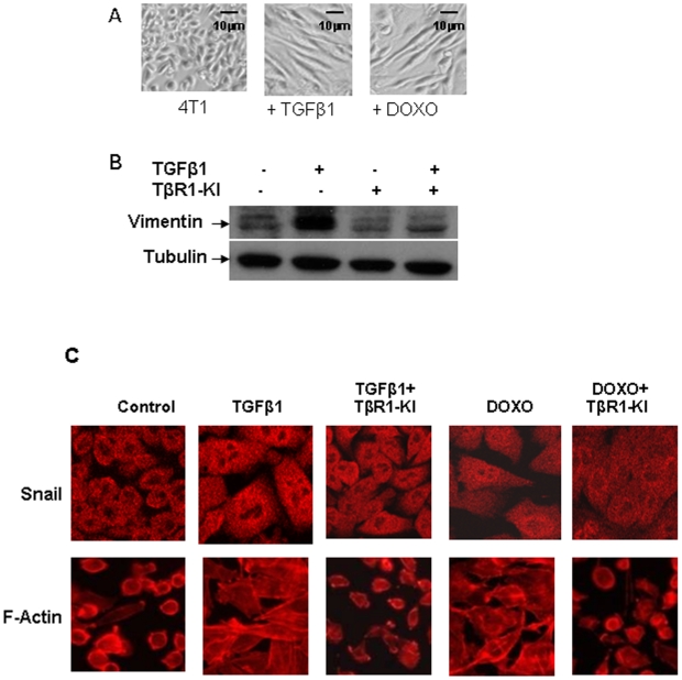
Background: Recent studies suggested that induction of epithelial-mesenchymal transition (EMT) might confer both metastatic and self-renewal properties to breast tumor cells resulting in drug resistance and tumor recurrence. TGFbeta is a potent inducer of EMT and has been shown to promote tumor progression in various breast cancer cell and animal models.

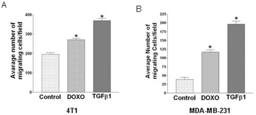
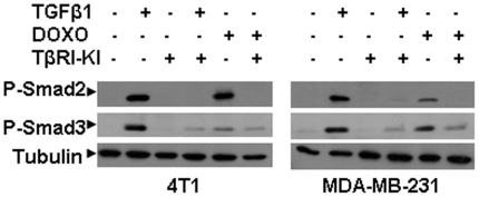
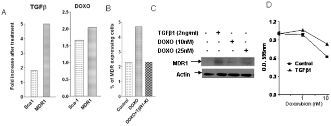
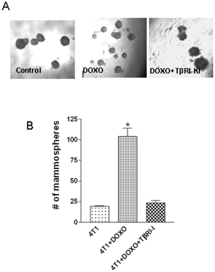
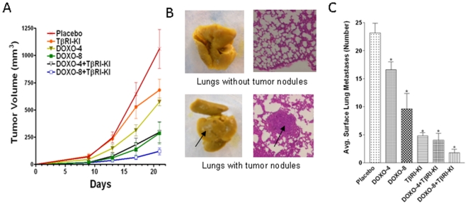
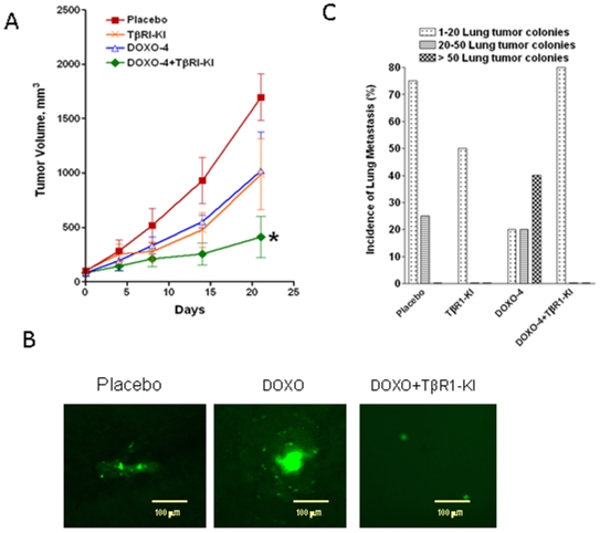
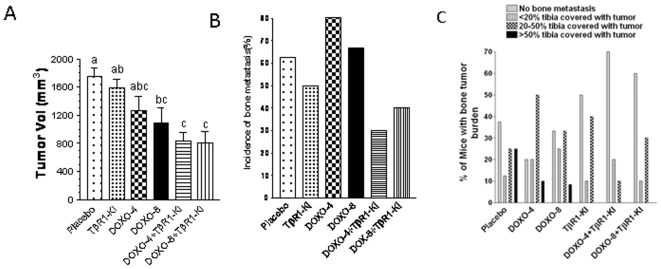
Principal findings: We report that chemotherapeutic drug doxorubicin activates TGFbeta signaling in human and murine breast cancer cells. Doxorubicin induced EMT, promoted invasion and enhanced generation of cells with stem cell phenotype in murine 4T1 breast cancer cells in vitro, which were significantly inhibited by a TGFbeta type I receptor kinase inhibitor (TbetaRI-KI). We investigated the potential synergistic anti-tumor activity of TbetaR1-KI in combination with doxorubicin in animal models of metastatic breast cancer. Combination of Doxorubicin and TbetaRI-KI enhanced the efficacy of doxorubicin in reducing tumor growth and lung metastasis in the 4T1 orthotopic xenograft model in comparison to single treatments. Doxorubicin treatment alone enhanced metastasis to lung in the human breast cancer MDA-MB-231 orthotopic xenograft model and metastasis to bone in the 4T1 orthotopic xenograft model, which was significantly blocked when TbetaR1-KI was administered in combination with doxorubicin.

Conclusions: These observations suggest that the adverse activation of TGFbeta pathway by chemotherapeutics in the cancer cells together with elevated TGFbeta levels in tumor microenvironment may lead to EMT and generation of cancer stem cells resulting in the resistance to the chemotherapy. Our results indicate that the combination treatment of doxorubicin with a TGFbeta inhibitor has the potential to reduce the dose and consequently the toxic side-effects of doxorubicin, and improve its efficacy in the inhibition of breast cancer growth and metastasis.


Conflict of interest statement

Competing Interests: The authors have declared that no competing interests exist.

Figures

Figure 1
Figure 1. Stimulation of migration by doxorubicin and TGFβ treatment in murine and human breast cancer cells.
In a Boyden Chamber migration assay, 40,000 murine breast cancer 4T1 cells (A) and human breast cancer MDA-MB-231 cells (B) in serum free medium were placed inside the insert of an invasive chamber and treated with doxorubicin (100 nM) or TGFβ1 (5 ng/ml in A and 1 ng/ml in B) for 6 hours (4T1 cells) and 24 hours (MDA-MB-231 cells). Lower chamber contained the medium with 1% serum as chemoattractant. Cells were removed from the upper surface of the chamber membrane and the membrane was then stained for the migratory cells, which were counted under a microscope. Results are the mean of the five fields of observation at 100× magnification and expressed as mean+SEM. “*” indicates significant difference from the control value with ANOVA test, P<0.05.
Figure 2
Figure 2. Enhanced phosphorylation of Smad2 and Smad3 in murine and human breast cancer cells by doxorubicin and TGFβ treatment.
The murine 4T1 and human MDA-MB-231 breast cancer cells were treated with doxorubicin (25 nM) or TGFβ1(2 ng/ml) in the presence or absence of TGFβ antagonist TbRI-KI (500 nM) for 7 days. The medium was changed every alternate day. The cell extracts were used for Western blotting analysis to measure the levels of phosphorylated Smad2 (P-Smad2) and phosphorylated Smad3 (P-Smad3) with their respective antibodies.
Figure 3
Figure 3. Inhibition of Doxorubicin- and TGFβ-induced EMT by TβRI-KI in murine 4T1 cells.
A. Representative pictures of 4T1 cells with mesenchymal morphology after treatment with TGFβ1 (2 ng/ml) or doxorubicin (25 nM) for 7 days (×100 magnification). B. 4T1 cells were treated with or without TGFβ1 (2 ng/ml) or TbRI-KI (500 nM) for 7 days. Cell lysates were analyzed by western blotting with antibody to Vimentin and tubulin. C. 4T1 cells were treated with TGFβ1 (2 ng/ml) or doxorubicin (25 nM) in the presence or absence of TbRI-KI (500 nM) for 7 days. The treated and non-treated 4T1 cells were harvested and about 4×10³ cells were seeded on a 8-well Lab-Tek chambered coverglass. After 24 hours, the cells were stained with Rhodamine phalloidin for F-actin or an antibody to Snail. Fluorescent images were taken with a confocal microscope.
Figure 4
Figure 4. Doxorubicin and TGFβ treatment generated drug resistant cells with a stem cell phenotype.
A and B. The murine 4T1 breast cancer cells were treated with TGFβ1 (2 ng/ml) or doxorubicin (25 nM) in the presence or absence of TbRI-KI (500 nM) for 7 days. The medium was changed every alternate day. The treated and non-treated cells were harvested with trypsinization and incubated with an alexafluor 405-conjugated antibody (2 µg/106 cells) against MDR1 or a Phycoerythrin-conjugated antibody against Sca-1. Fluorescence Activated Cell Sorting (FACS) analysis was carried out using FACSAria flow cytometer (Becton Dickinson). Results were expressed as fold increase of the number of cells expressing the surface markers for Sca-1 or MDR1 after treatment with TGFβ1 or doxorubicin compared to the untreated cells (A) or as the percentage of MDR1 expressing cells after doxorubicin treatment in the presence or absence of TβR1-KI (B). C. 4T1 cells were treated with or without 2 ng/ml TGFβ1 and DOXO (10 nM and 25 nM) for 7 days and cell extracts were used for Western blot analysis with antibody to MDR1. D. 4T1 cells, pre-treated with TGFβ1 (5 ng/ml) for seven days, were plated in a 96-well plate and were treated with doxorubicin at two different concentrations (1 nM and 10 nM) in the presence or absence of TGFβ1 (5 ng/ml). Relative cell number after 5 days of incubation was obtained by MTT assay. Each data point is the mean±SEM from 4 replicate wells.
Figure 5
Figure 5. Abrogation of doxorubicin induced mammosphere formation by TβRI-KI.
Murine 4T1 cells were treated with doxorubicin (25 nM) in the presence or absence of TbRI-KI for 6 days. The cells with or without treatment were harvested after trypsinization and filtered through 40 µm strainer to obtain a single-cell suspension. Cells were seeded (1,000 cells/100 µl/well) in a 96-well ultra-low attachment plate (Costar) and mammospheres were counted after 7 days of incubation. A. Representative pictures of mammospheres obtained from cells with or without doxorubicin treatment and also after doxorubicin treatment in the presence of TbRI-KI. B. The number of mammospheres obtained from 1,000 cells in triplicate wells with or without treatments were counted and expressed as mean±SEM. “*” indicates significant difference from the control value, P<0.05.
Figure 6
Figure 6. TGFβ inhibitor enhances the efficacy of doxorubicin treatment to reduce tumor growth and spontaneous lung metastasis of murine breast cancer cells.
A. Murine breast cancer 4T1 cells (0.1×10⁶) were inoculated at the both inguinal mammary fat pad of syngeneic Balb/c mice and divided into six groups. Animals were treated with PBS (Placebo), TβRI-KI at 1 mg/kg every alternative day, doxorubicin (DOXO) at 4 mg/kg (DOXO-4) or 8 mg/kg (DOXO-8) once per 7 days, or the combination of TβRI-KI and DOXO-4 or DOXO-8 through i.v. injection starting at the day of tumor cell inoculation. Tumor sizes were measured twice weekly with a caliper, and the tumor volumes were calculated with the equation V = (L×W2)×0.5, where L is length and W is width of a tumor. Experiment was terminated after 3 wks of treatment and the final tumor volume (sum of two tumors per mouse) was used for calculating mean tumor volume ± SEM from 6 mice in Placebo and 5 mice in the rest of the groups. B and C. After the termination of the experiment, lungs were excised and fixed in Bouin's solution (Sigma) to count surface lung metastasis, followed by paraffin embedding for histological H & E staining. Representative picture of lungs without and with metastatic nodules are shown with corresponding histological identification of the nodules by H & E staining (B). The visible surface lung nodules were counted. “*” denotes a significant difference (P<0.005) from the corresponding control with a Student t test (C).
Figure 7
Figure 7. Treatment with TβRI-KI blocked doxorubicin-induced spontaneous lung metastasis of human breast cancer cells.
A. EGFP-expressing sub confluent MDA-MB-231 cells (1×10⁶) were inoculated into both inguinal mammary fat pads of female athymic nude mice. Tumor volume was measured externally and calculated as described in the legend of Figure 5. Tumors were allowed to reach 80–100 mm3 in size and the animals were then ranked according to their tumor burden and divided into four groups such that the mean and the median of the tumor volume of the groups are closely matched. Animals were treated with PBS (control), TβRI-KI every alternative day, doxorubicin once every week, and with the combination of TβRI-KI and doxorubicin respectively. The experiment was terminated after 3 weeks and the final tumor volume (sum of two tumors per mouse) was used for calculating mean tumor volume±SEM from 4 mice in control and TβRI-KI groups and 5 mice in the other two groups. Asterisk “*” denotes significant difference (P<0.05) of the terminal tumor volumes between control and the combination treatment group by ANOVA. B. Representative Pictures of lung metastasis colonies formed by EGFP-expressing MDA-MB-231 cells detected by fluorescence microscopy (×200 magnification). C. Green fluorescent micrometastatic colonies in the whole lung were detected and counted. The percent of mice with lung metastasis in each group is expressed as total lung metastasis incidence. Incidence as percent of mice with numbers of lung metastasis colonies ranging between 1 and 20, between 20 and 50, and >50 is also presented.
Figure 8
Figure 8. TβRI-KI reduced doxorubicin-induced spontaneous bone metastasis in the murine xenograft model.
A. 4T1 cells (0.1×10⁶) were inoculated into both inguinal mammary fat pads of syngeneic female balb/c mice. When the average tumor volume reached 60–70 mm3 after 2 weeks, the mice were divided into six groups with similar tumor burden and treated i.p. with PBS (control), TβRI-KI at 1 mg/kg every alternative day, doxorubicin at 4 mg/kg (DOXO-4) or 8 mg/kg (DOXO-8) once per 7 days, or the combination of TβRI-KI and DOXO-4 or DOXO-8. The experiment was terminated after 3 wks of treatment and the final tumor volume (sum of two tumors per mouse) was used for calculating mean tumor volume±SEM from 6 mice in DOXO-8 and 5 mice in the rest groups. The bars bearing a different letter are significantly different (P<0.05) by ANOVA. B. After the termination of the experiment, the leg bones (Femora and tibiae) were fixed in 10% neutral buffered formalin, decalcified in 10% EDTA and embedded in paraffin. Sections were stained with H&E, orange G and phloxine. The presence of tumors in the bone marrow was examined microscopically. The incidence of bone metastasis in each group of animals was expressed in percentage of the number of animals with bone metastasis. C. Animals with bone tumor burden were divided into four groups and the results were expressed in percentage of animals with no bone metastasis, with <20% tibia replaced by tumor, with 20–50% tibia replaced by tumor, and with >50% of tibia replaced by tumor.

Similar articles

Cited by

References

    1. Parkin DM, Bray F, Ferlay J, Pisani P. Global cancer statistics, 2002. CA Cancer J Clin. 2005;55:74–108. - PubMed
    1. Gamucci T, D'Ottavio AM, Magnolfi E, Barduagni M, Vaccaro A, et al. Weekly epirubicin plus docetaxel as first-line treatment in metastatic breast cancer. Br J Cancer. 2007;97:1040–1045. - PMC - PubMed
    1. Sledge GW, Neuberg D, Bernardo P, Ingle JN, Martino S, et al. Phase III trial of doxorubicin, paclitaxel, and the combination of doxorubicin and paclitaxel as front-line chemotherapy for metastatic breast cancer: an intergroup trial (E1193). J Clin Oncol. 2003;21:588–592. - PubMed
    1. Ellis MJ, Hayes DF, Lippman ML. Treatment of metastatic breast cancer: Diseases of the breast. 2004;3rd:749–797.
    1. Chen JH, Ling R, Yao Q, Li Y, Chen T, et al. Effect of small-sized liposomal Adriamycin administered by various routes on a metastatic breast cancer model. Endocr Relat Cancer. 2005;12:93–100. - PubMed

Publication types

MeSH terms

Substances