

Keratinocytes determine Th1 immunity during early experimental leishmaniasis
- PMID: 20442861
- PMCID: PMC2861693
- DOI: 10.1371/journal.ppat.1000871
Keratinocytes determine Th1 immunity during early experimental leishmaniasis
Abstract
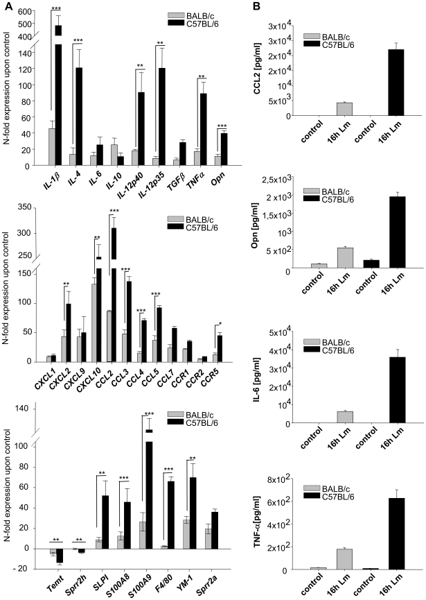
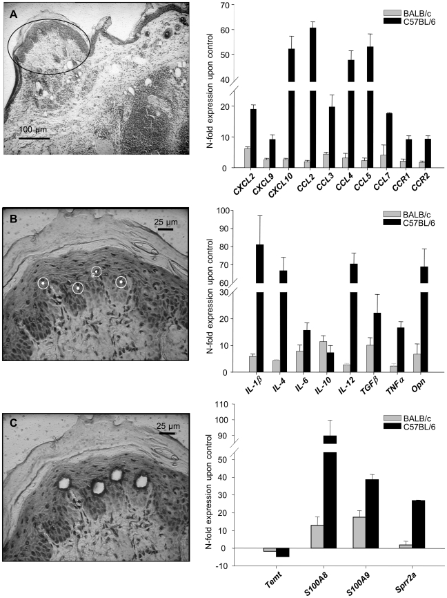
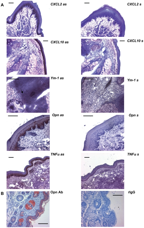
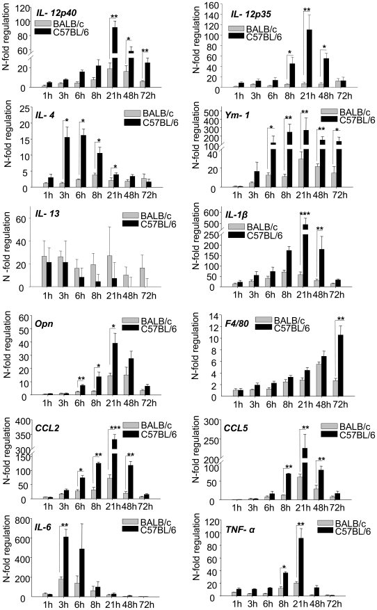
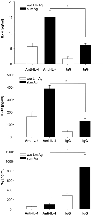
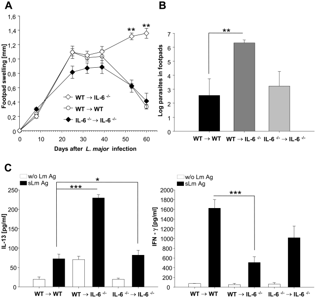
Experimental leishmaniasis is an excellent model system for analyzing Th1/Th2 differentiation. Resistance to Leishmania (L.) major depends on the development of a L. major specific Th1 response, while Th2 differentiation results in susceptibility. There is growing evidence that the microenvironment of the early affected tissue delivers the initial triggers for Th-cell differentiation. To analyze this we studied differential gene expression in infected skin of resistant and susceptible mice 16h after parasite inoculation. Employing microarray technology, bioinformatics, laser-microdissection and in-situ-hybridization we found that the epidermis was the major source of immunomodulatory mediators. This epidermal gene induction was significantly stronger in resistant mice especially for several genes known to promote Th1 differentiation (IL-12, IL-1beta, osteopontin, IL-4) and for IL-6. Expression of these cytokines was temporally restricted to the crucial time of Th1/2 differentiation. Moreover, we revealed a stronger epidermal up-regulation of IL-6 in the epidermis of resistant mice. Accordingly, early local neutralization of IL-4 in resistant mice resulted in a Th2 switch and mice with a selective IL-6 deficiency in non-hematopoietic cells showed a Th2 switch and dramatic deterioration of disease. Thus, our data indicate for the first time that epidermal cytokine expression is a decisive factor in the generation of protective Th1 immunity and contributes to the outcome of infection with this important human pathogen.
Conflict of interest statement
The authors have declared that no competing interests exist.
Figures
Similar articles
-
Epidermal expression of I-TAC (Cxcl11) instructs adaptive Th2-type immunity.FASEB J. 2014 Apr;28(4):1724-34. doi: 10.1096/fj.13-233593. Epub 2014 Jan 7. FASEB J. 2014. PMID: 24398292
-
The mechanism of in vitro T helper cell type 1 to T helper cell type 2 switching in highly polarized Leishmania major-specific T cell populations.J Immunol. 1997 Feb 15;158(4):1559-64. J Immunol. 1997. PMID: 9029090
-
Interleukin 1alpha promotes Th1 differentiation and inhibits disease progression in Leishmania major-susceptible BALB/c mice.J Exp Med. 2003 Jul 21;198(2):191-9. doi: 10.1084/jem.20030159. Epub 2003 Jul 14. J Exp Med. 2003. PMID: 12860932 Free PMC article.
-
Anti-leishmania effector functions of CD4+ Th1 cells and early events instructing Th2 cell development and susceptibility to Leishmania major in BALB/c mice.Adv Exp Med Biol. 1998;452:53-60. doi: 10.1007/978-1-4615-5355-7_7. Adv Exp Med Biol. 1998. PMID: 9889959 Review.
-
The Th1/Th2 paradigm and experimental murine leishmaniasis.Int Arch Allergy Immunol. 1998 Mar;115(3):191-202. doi: 10.1159/000023900. Int Arch Allergy Immunol. 1998. PMID: 9531160 Review.
Cited by
-
Early Leukocyte Responses in Ex-Vivo Models of Healing and Non-Healing Human Leishmania (Viannia) panamensis Infections.Front Cell Infect Microbiol. 2021 Sep 7;11:687607. doi: 10.3389/fcimb.2021.687607. eCollection 2021. Front Cell Infect Microbiol. 2021. PMID: 34557423 Free PMC article.
-
Concurrent exposure to a dectin-1 agonist suppresses the Th2 response to epicutaneously introduced antigen in mice.J Biomed Sci. 2013 Jan 3;20(1):1. doi: 10.1186/1423-0127-20-1. J Biomed Sci. 2013. PMID: 23286586 Free PMC article.
-
Cutaneous Manifestations of Human and Murine Leishmaniasis.Int J Mol Sci. 2017 Jun 18;18(6):1296. doi: 10.3390/ijms18061296. Int J Mol Sci. 2017. PMID: 28629171 Free PMC article. Review.
-
Deletion of Interleukin-4 Receptor Alpha-Responsive Keratinocytes in BALB/c Mice Does Not Alter Susceptibility to Cutaneous Leishmaniasis.Infect Immun. 2018 Nov 20;86(12):e00710-18. doi: 10.1128/IAI.00710-18. Print 2018 Dec. Infect Immun. 2018. PMID: 30275010 Free PMC article.
-
TLR2 Signaling in Skin Nonhematopoietic Cells Induces Early Neutrophil Recruitment in Response to Leishmania major Infection.J Invest Dermatol. 2019 Jun;139(6):1318-1328. doi: 10.1016/j.jid.2018.12.012. Epub 2018 Dec 27. J Invest Dermatol. 2019. PMID: 30594488 Free PMC article.
References
-
- Sacks D, Noben-Trauth N. The immunology of susceptibility and resistance to Leishmania major in mice. Nat Rev Immunol. 2002;2:845–858. - PubMed
-
- Sunderkotter C, Kunz M, Steinbrink K, Meinardus-Hager G, Goebeler M, et al. Resistance of mice to experimental leishmaniasis is associated with more rapid appearance of mature macrophages in vitro and in vivo. J Immunol. 1993;151:4891–4901. - PubMed
-
- Tacchini-Cottier F, Zweifel C, Belkaid Y, Mukankundiye C, Vasei M, et al. An immunomodulatory function for neutrophils during the induction of a CD4+ Th2 response in BALB/c mice infected with Leishmania major. J Immunol. 2000;165:2628–2636. - PubMed
-
- Nabors GS, Nolan T, Croop W, Li J, Farrell JP. The influence of the site of parasite inoculation on the development of Th1 and Th2 type immune responses in (BALB/c×C57BL/6) F1 mice infected with Leishmania major. Parasite Immunol. 1995;17:569–579. - PubMed
Publication types
MeSH terms
Substances
LinkOut - more resources
Full Text Sources
Other Literature Sources
Research Materials
