Review
. 2010 May;13(3):335-49.

Advances in protein NMR provided by the NIGMS Protein Structure Initiative: impact on drug discovery

Affiliations
Review

Advances in protein NMR provided by the NIGMS Protein Structure Initiative: impact on drug discovery

Gaetano T Montelione et al. Curr Opin Drug Discov Devel. 2010 May.

Abstract

Rational drug design relies on the 3D structures of biological macromolecules, with a particular emphasis on proteins. The structural genomics-based high-throughput structure determination platforms established by the Protein Structure Initiative (PSI) of the National Institute of General Medical Science (NIGMS) of the NIH are uniquely suited to provide these structures. NMR plays a critical role in structure determination because many important protein targets do not form the single crystals required for X-ray diffraction. NMR can provide valuable structural and dynamic information on proteins and their drug complexes that cannot be obtained with X-ray crystallography. This review discusses recent advances in NMR that have been driven by structural genomics projects. These advances suggest that the future discovery and design of drugs can increasingly rely on protocols using NMR approaches for the rapid and accurate determination of structures.


Figures

Fig. 1
Fig. 1
Three protein-ligand complexes solved using NMR by the NESG. Proteins are shown as ribbon draws and ligands are shown as sticks (a) ZR31, Staphylococcus aureus SACOL2532 with CoA (PDB code 2h5m and 1r57); (b) ET106, E. coli YhhK with CoA, (PDB code 2k5t); (c) GmR141, Geobacter metallireducens Gmet_2339 with phosphopanthethine, (PDB code 2kjs ). These structures of protein-ligand complexes were determined by J. Cort, T. Ramelot, and M. Kennedy.
Fig. 2
Fig. 2
(A,B) NMR structure production of NESG (green), CESG (blue), and JCSG (red) PSI Centers. (C) MW distribution of NMR structures deposited by nonSG (black) and PSI SG (green) groups since 2000. Distribution in MW of NMR structures deposited by PSI groups (which is dominated by structures determined by the NESG Consortium. The PSI NMR structures have a mean MW of ~ 13 kDa, similar to that of NMR structures deposited in the PDB by nonSG groups (12 kDa, excluding peptides of < 4,000 Da) over the last 10 years.
Fig. 3
Fig. 3
Cumulative PDB depositions of NESG NMR and X-ray crystal structures in each month of the PSI Program.
Fig. 4
Fig. 4
MolProbity packing [28] Z scores for NMR structures deposited in PDB by NESG over the first 9 years of PSI (A) demonstrate significantly better scores than for NMR structures for a random set of nonSG structures of similar size deposited in the PDB during the same time period. Average Z score indicated by red dotted line. Similar conclusions are reached looking at sidechain dihedral angle distributions.
Fig. 5
Fig. 5
Plots of ProCheck_(bb+sc dihedral) vs. MolProbity Z scores demonstrate higher quality of PSI (and particularly NESG) NMR structures than for nonSG structures. Z scores are normalized to Z=0 for average values of quality scores obtained using a set of medium-sized (100 – 500 residue) high-resolution (1.0 - 1.8 Å) crystal structures [26]. A large number of nonSG NMR structures with Z scores < -20 are not shown in the plots.
Fig. 6
Fig. 6
HDX-MS-based construct optimization [58] of E. coli yiaD (NESG target, ER553). (A) 2D [1H-15N] HSQC spectra of full length (left) and construct optimized (right) E. coli yiaD. (B) HDX-MS data for full-length E. coli yiaD (rows show degree of exchange obtained with 10, 100, and 1000 second 1H/2H exchange durations at 4 °C). The results are depicted in a so-called “heat map”, in which 1H/2H exchange rates are represented by colors ranging from blue (slow amide proton exchange) to red (fast amide proton exchange). The NMR structure of a HDX-MS optimized construct of ER553 was subsequently solved (PDB_ID 2k1s).
Fig. 7
Fig. 7
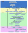
Flow-chart of NESG NMR structure production comprising (from top to bottom) sample preparation, NMR data collection, structure calculation, and structure validation and deposition. For details of the associated protocols see ref. 95.
Fig. 8
Fig. 8
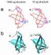
Assessment of structural accuracy for an NMR protein structure determined with a 600 MHz 1-mm micro NMR probe [89]. (A) Backbone superimposition and (B) ribbon diagrams of the solution NMR structures of NESG target MaR30 determined using conventional (left) and microprobe (right) data. The backbone root mean squared deviation (rmsd) between mean coordinates of the ensembles of conventional and microprobe structures is 0.7 Å, demonstrating that high-quality structures can be obtained with < 100 ug protein samples. Several other protein NESG protein structures have been determined using even higher quality data obtained with a 600 MHz 1.7-mm cryo microprobe.

Similar articles

Cited by

References

    1. Liu J, Montelione GT, Rost B. Novel leverage of structural genomics. Nat Biotechnol. 2007;25:849–851. Describes the metric of “novel structural leverage” and an assessment of the impact of SG efforts in coverage of protein sequences with representative 3D structures.

    1. Nair R, Liu J, Soong TT, Acton TB, Everett JK, Kouranov A, Fiser A, Godzik A, Jaroszewski L, Orengo C, et al. Structural genomics is the largest contributor of novel structural leverage. J Struct Funct Genomics. 2009;10:181–191. - PMC - PubMed
    1. Schwede T, Sali A, Honig B, Levitt M, Berman HM, Jones D, Brenner SE, Burley SK, Das R, Dokholyan NV, et al. Outcome of a workshop on applications of protein models in biomedical research. Structure. 2009;17:151–159. - PMC - PubMed
    1. Dessailly BH, Nair R, Jaroszewski L, Fajardo JE, Kouranov A, Lee D, Fiser A, Godzik A, Rost B, Orengo C. PSI-2: structural genomics to cover protein domain family space. Structure. 2009;17:869–881. Description of the targeting strategy of the PSI program for broad structural coverage, it rational, and progress, written by the group responsible for selecting these domain families.

    1. Levitt M. Nature of the protein universe. Proc Natl Acad Sci U S A. 2009;106:11079–11084. Assessment of the numbers of domain families indicated by currently available protein sequence data, suggesting that the majority of protein sequences can be clustered into some 15,000 - 20,000 domain families.

Publication types

MeSH terms

LinkOut - more resources