. 2010 May;11(5):307-14.
doi: 10.1631/jzus.B0900391.

Morphologic observation and classification criteria of atretic follicles in guinea pigs

Affiliations

Morphologic observation and classification criteria of atretic follicles in guinea pigs

Wei Wang et al. J Zhejiang Univ Sci B. 2010 May.

Abstract

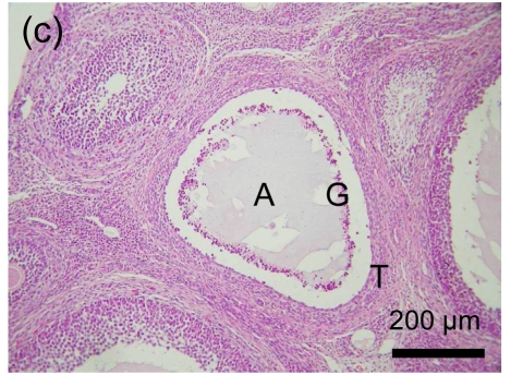
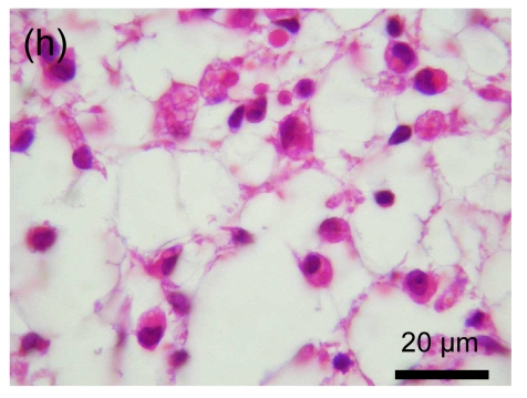
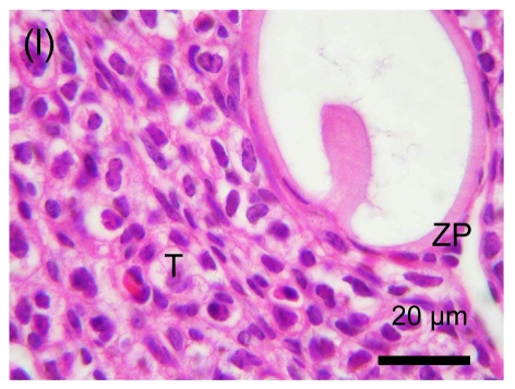
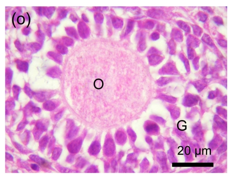
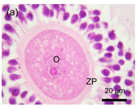
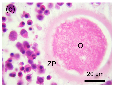
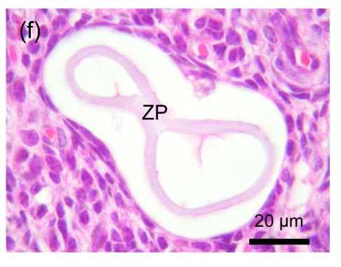
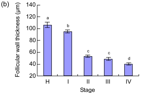
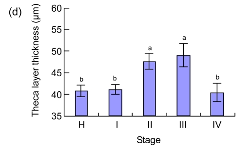
There is a lack of appropriate classification criteria for the determination of atretic follicles in guinea pigs. In the present study, new criteria were established based on the latest morphologic criteria for cell death proposed by the Nomenclature Committee on Cell Death (NCCD) in 2009. Ovaries of guinea pigs were sampled on different stages of estrous cycle, and the morphologic observations of atretic follicles were investigated in serial sections. The results showed that the process of follicular atresia could be classified into four continuous stages: (1) the granulosa layer became loose, and some apoptotic bodies began to appear; (2) the granulosa cells were massively eliminated; (3) the theca interna cells differentiated; and (4) the residual follicular cells degenerated. In addition, the examination revealed that these morphologic criteria were accurate and feasible. In conclusion, this study provides new criteria for the classification of atretic follicles in guinea pigs, and this knowledge can inform future research in the area.


Figures

Fig. 1
Fig. 1
Morphologic observation of follicular atresia (a) & (b) Stage I: granulosa layer became loose and the pyknotic cells appeared; (c), (d) & (e) Stage II: granulosa cells were eliminated massively and floated in the antrum; (f), (g) & (h) Early phase of Stage III: atrophic follicular cavity was occupied by fibroblast-like cells with slender shapes (g) and round shapes (h); (i) & (j) Late phase of Stage III: at first, only a small quantity of differentiated cells appeared around the zona pellucida (i), however, it increased greatly and formed a new tissue subsequently (j); (k) & (l) Stage IV: follicle volume became smaller and smaller; (m) & (n) Abnormal manner of follicular atresia: in some small antral follicles (m), the cumulus granulosa cells were eliminated first and the granulosa layers were relatively intact (n); (o) & (p) Preantral follicular atresia: there was no massive elimination of cells (o), preantral follicles degenerated gradually (p). O: oocyte; G: granulosa layer; T: theca layer; A: follicular antrum; C: Call-Exner body; ZP: zona pellucida
Fig. 1
Fig. 1
Morphologic observation of follicular atresia (a) & (b) Stage I: granulosa layer became loose and the pyknotic cells appeared; (c), (d) & (e) Stage II: granulosa cells were eliminated massively and floated in the antrum; (f), (g) & (h) Early phase of Stage III: atrophic follicular cavity was occupied by fibroblast-like cells with slender shapes (g) and round shapes (h); (i) & (j) Late phase of Stage III: at first, only a small quantity of differentiated cells appeared around the zona pellucida (i), however, it increased greatly and formed a new tissue subsequently (j); (k) & (l) Stage IV: follicle volume became smaller and smaller; (m) & (n) Abnormal manner of follicular atresia: in some small antral follicles (m), the cumulus granulosa cells were eliminated first and the granulosa layers were relatively intact (n); (o) & (p) Preantral follicular atresia: there was no massive elimination of cells (o), preantral follicles degenerated gradually (p). O: oocyte; G: granulosa layer; T: theca layer; A: follicular antrum; C: Call-Exner body; ZP: zona pellucida
Fig. 2
Fig. 2
Morphologic observation of oocyte degeneration (a) & (b) Stage I. Some cavities appeared in the oocyte; (c) Stage II. The oocyte shrank to a certain extent, and the cumulus granulosa cells receded from the zona pellucida; (d) Early phase of Stage III. The characteristics of degeneration were obvious; (e) Late phase of Stage III. The oocyte degenerated to a small volume; (f) Stage IV. The oocyte degenerated completely. O: oocyte; ZP: zona pellucida
Fig. 2
Fig. 2
Morphologic observation of oocyte degeneration (a) & (b) Stage I. Some cavities appeared in the oocyte; (c) Stage II. The oocyte shrank to a certain extent, and the cumulus granulosa cells receded from the zona pellucida; (d) Early phase of Stage III. The characteristics of degeneration were obvious; (e) Late phase of Stage III. The oocyte degenerated to a small volume; (f) Stage IV. The oocyte degenerated completely. O: oocyte; ZP: zona pellucida
Fig. 3
Fig. 3
Measurements of the follicular diameter (a) and thicknesses of follicular wall (b), granulosa layer (c) and theca layer (d) Measurements were taken of healthy follicles (H, n=28) and atresia follicles in Stages I (n=46), II (n=35), III (n=37) and IV (n=33). Each value represents the mean±SEM. One-way analysis of variance (ANOVA) was used to analyze the data. a, b, c, d Significant values (P<0.05) by Tukey’s multiple-range test
Fig. 3
Fig. 3
Measurements of the follicular diameter (a) and thicknesses of follicular wall (b), granulosa layer (c) and theca layer (d) Measurements were taken of healthy follicles (H, n=28) and atresia follicles in Stages I (n=46), II (n=35), III (n=37) and IV (n=33). Each value represents the mean±SEM. One-way analysis of variance (ANOVA) was used to analyze the data. a, b, c, d Significant values (P<0.05) by Tukey’s multiple-range test

Similar articles

Cited by

References

    1. Bland KP. Biphasic follicular growth in the guinea-pig oestrous cycle. J Reprod Fertil. 1980;60(1):73–76. doi: 10.1530/jrf.0.0600073. - DOI - PubMed
    1. Byskov AG. Cell kinetic studies of follicular atresia in the mouse ovary. J Reprod Fertil. 1974;37(2):277–285. doi: 10.1530/jrf.0.0370277. - DOI - PubMed
    1. Cui Y, Yu SJ. Ovarian morphology and follicular systems in yaks of different ages. Vet J. 1999;157(2):197–205. doi: 10.1053/tvjl.1998.0282. - DOI - PubMed
    1. Fortune JE. Ovarian follicular growth and development in mammals. Biol Reprod. 1994;50(2):225–232. doi: 10.1095/biolreprod50.2.225. - DOI - PubMed
    1. Galluzzi L, Maiuri MC, Vitale I, Zischka H, Castedo M, Zitvogel L, Kroemer G. Cell death modalities: classification and pathophysiological implications. Cell Death Differ. 2007;14(7):1237–1243. doi: 10.1038/sj.cdd.4402148. - DOI - PubMed

Publication types