. 2008 May;1(1):80-6.
doi: 10.1111/j.1752-8062.2008.00030.x.

The human heart: a self-renewing organ

Affiliations

The human heart: a self-renewing organ

Jan Kajstura et al. Clin Transl Sci. 2008 May.

Abstract

The dogma that the heart is a static organ which contains an irreplaceable population of cardiomyocytes prevailed in the cardiovascular field for the last several decades. However, the recent identification of progenitor cells that give rise to differentiated myocytes has prompted a re-interpretation of cardiac biology. The heart cannot be viewed any longer as a postmitotic organ characterized by a predetermined number of myocytes that is defined at birth and is preserved throughout life. The myocardium constitutes a dynamic entity in which new young parenchymal cells are formed to substitute old damaged dying myocytes. The regenerative ability of the heart was initially documented with a classic morphometric approach and more recently with the demonstration that DNA synthesis, mitosis, and cytokinesis take place in the newly formed myocytes of the normal and pathologic heart. Importantly, replicating myocytes correspond to the differentiated progeny of cardiac stem cells. These findings point to the possibility of novel therapeutic strategies for the diseased heart.


Figures

Figure 1
Figure 1
Cardiac hypertrophy and myocyte size. Section of a normal (A, heart weight = 220 g) and hypertrophied (B, heart weight = 700 g) human myocardium. The boundary of myocytes is defined by laminin (green). Note that the myocyte cross‐sectional area in both preparations is comparable, suggesting the presence of a higher number of myocytes in the heavier heart α‐SA, α‐sarcomeric actin (red).
Figure 2
Figure 2
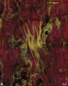
Myocardial damage. Panels A–D represent various aspects of myocardial fibrosis. Replacement fibrosis is recognized by foci of accumulation of collagen type I and type III (yellow) in hearts affected by chronic ischemia (A and B). Interstitial fibrosis is documented by the presence of collagen in the interstitial space between neighboring myocytes (C and D). Panel E illustrates a myocyte undergoing necrosis. Blunt‐end DNA strand breaks, typical of this type of cell death, are shown by in situ ligation of a Pfu oligonucleotide probe (yellow, arrow). Additionally, loss of plasmamembrane integrity (arrowheads) is visualized by a discontinuous vinculin staining (green). Panel E: modified from Ref. 26.
Figure 2
Figure 2
Myocardial damage. Panels A–D represent various aspects of myocardial fibrosis. Replacement fibrosis is recognized by foci of accumulation of collagen type I and type III (yellow) in hearts affected by chronic ischemia (A and B). Interstitial fibrosis is documented by the presence of collagen in the interstitial space between neighboring myocytes (C and D). Panel E illustrates a myocyte undergoing necrosis. Blunt‐end DNA strand breaks, typical of this type of cell death, are shown by in situ ligation of a Pfu oligonucleotide probe (yellow, arrow). Additionally, loss of plasmamembrane integrity (arrowheads) is visualized by a discontinuous vinculin staining (green). Panel E: modified from Ref. 26.
Figure 3
Figure 3
Myocyte proliferation. Myocyte generation in the diseased human heart. Metaphase chromosomes (arrow) are apparent in the dividing cardiomyocyte. Laminin (green) marks the boundaries between cells. Note the small size of the mitotic myocyte. Reprinted from Ref. 31.
Figure 4
Figure 4
Clonogenicity of human cardiac stem cells. (A) Growth of a clone from a single c‐kit positive cell over a period of 9 days (d). The lower right panel documents that cells in the clone retain the c‐kit antigen (green). (B) Higher magnification of a larger clone of human cardiac stem cells derived from a single c‐kit positive cell. Panel A: reprinted from Ref. 96.
Figure 4
Figure 4
Clonogenicity of human cardiac stem cells. (A) Growth of a clone from a single c‐kit positive cell over a period of 9 days (d). The lower right panel documents that cells in the clone retain the c‐kit antigen (green). (B) Higher magnification of a larger clone of human cardiac stem cells derived from a single c‐kit positive cell. Panel A: reprinted from Ref. 96.

Similar articles

Cited by

References

    1. Anversa P, Kajstura J. Ventricular myocytes are not terminally differentiated in the adult mammalian heart. Circ Res. 1998; 83: 1–14. - PubMed
    1. Karsner HT, Saphir O, Todd TW. The state of the cardiac muscle in hypertrophy and atrophy. Am J Pathol. 1925; 1: 351–371. - PMC - PubMed
    1. Petersen RO, Baserga R. Nucleic acid and protein synthesis in cardiac muscle of growing and adult mice. Exp Cell Res. 1965; 40: 340–352. - PubMed
    1. Morkin E, Ashford TP. Myocardial DNA synthesis in experimental cardiac hypertrophy. Am J Physiol. 1968; 215: 1409–1413. - PubMed
    1. Grove D, Nair KG, Zak R. Biochemical correlates of cardiac hypertrophy, III: changes in DNA content: the relative contributions of polyploidy and mitotic activity. Circ Res. 1969; 25: 463–471. - PubMed

Publication types

LinkOut - more resources