

doi: 10.1111/j.1752-8062.2008.00037.x.
Overexpression of matrix metalloproteinase 9 in tumor epithelial cells correlates with colorectal cancer metastasis
Affiliations
- PMID: 20443834
- PMCID: PMC5439552
- DOI: 10.1111/j.1752-8062.2008.00037.x
Item in Clipboard
Overexpression of matrix metalloproteinase 9 in tumor epithelial cells correlates with colorectal cancer metastasis
Clin Transl Sci.
2008 Sep.
Abstract
Colorectal cancer mortality largely reflects metastasis, the spread of the disease to distant organs. Matrix metalloproteinase 9 (MMP-9) is a key regulator of metastasis and a target for anticancer strategies in colon cancer. Here, the overexpression of MMP-9 in pure tumor epithelial, but nor stromal, cell populations was associated with metastatic progression of colorectal cancer, as defined by reverse transcriptase-polymerase chain reaction (qRT-PCR) and confirmed by immunostaining. Thus, cancer cell MMP-9 represents a novel, selective prognostic and predictive factor that may be exploited for more effective disease stage stratification and therapeutic regimen selection in patients with colorectal cancer.
Figures
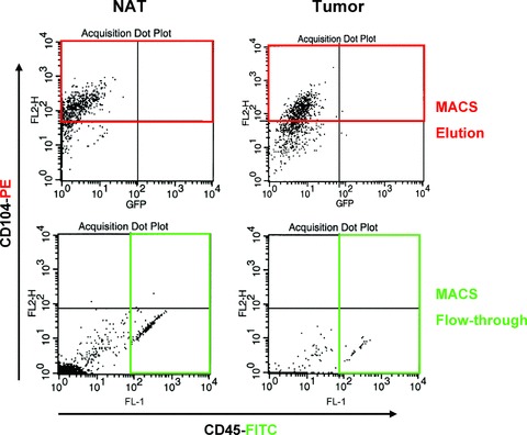
Isolation of pure epithelial and stromal cell populations from primary tumors and matched normal mucosa of patients with colorectal cancer. Following collagenase digestion and MACS sorting, the epithelial (MACS elution) or stromal (MACS flow‐through) cells of normal adjacent tissue (NAT) and colorectal tumors (Tumor) isolated from patients were subjected to FACS sorting. Pure intestinal epithelial (red box for PE‐conjugated anti‐CD104) or stromal (green box for FITC‐conjugated anti‐CD45) cell populations were collected for further analysis.

Relative MMP‐9 expression levels in colorectal tumor cell populations exhibit prognostic significance. (A) Stromal and epithelial cells from primary colorectal tumors (Tumor) and normal adjacent tissue (NAT) isolated by MACS and FACS sorting were subjected to RT‐PCR. Sample levels of MMP‐9 mRNA were normalized to respective β‐actin mRNA using the formula 2[(MMP‐9
Ct
) −(β‐actin
Ct
)], where Ct is the sample threshold cycle number. The lines connect data from matched specimens from the same patient. The box plots denote median and values from the 25th to 75th percentile; the whiskers denote range of values. (B) Lymph nodes involvement in the different subgroups of patients as defined by the relative expression of tumor cell MMP‐9 compared to the matched normal mucosa cell MMP‐9. ↑MMP‐9 = MMP‐9 overexpression; ↓MMP‐9 = MMP‐9 downregulation; E = tumor epithelial cells; S = tumor stromal cells.

MMP‐9 overexpression in tumor epithelial cells is a biological marker of colorectal cancer metastasis. Association between MMP‐9 overexpression in tumor cell populations and lymph node involvement (left panels). Compared to those with tumor MMP‐9 downregulation, patients with MMP‐9 overexpression in tumor epithelial, but not stromal, cells exhibit advanced disease stage (middle panels) and increased lymph node tumor burden (right panels). N categories and disease stage scores are specified in
Table 2
and are based on the AJCC/TNM system.
19
↑MMP‐9 = MMP‐9 overexpression; ↓MMP‐9 = MMP‐9 downregulation. The values in the middle and right panels are means ± SEM. *p <0.05 by the Wilcoxon two‐sample test.

Overexpression of tumor epithelial cell MMP‐9 in metastatic colorectal cancer is detected by immunostaining. IHC (left panels) and immunofluorescence (right panels) analyses of primary tumors (Tumor) and matched normal adjacent tissues (NAT) from colorectal cancer patients with metastatic disease progression. For IHC (magnification 40×), the tissues were stained with specific goat polyclonal anti‐MMP‐9 (brown) and hematoxylin (blue, nuclei). For immunofluorescence, the tissues were stained with DAPI (blue, nuclei) and specific antibodies against MMP‐9 (red) and the epithelial‐specific marker β‐catenin (green) and subjected to confocal microscopy (magnification 100×).
Similar articles
-
Tumor epithelial cell matrix metalloproteinase 9 is a target for antimetastatic therapy in colorectal cancer.Clin Cancer Res. 2006 Mar 15;12(6):1876-82. doi: 10.1158/1078-0432.CCR-05-2686. Clin Cancer Res. 2006. PMID: 16551873
-
Overexpression of MMP-13 gene in colorectal cancer with liver metastasis.Anticancer Res. 2010 Jul;30(7):2693-9. Anticancer Res. 2010. PMID: 20683000
-
The membrane-anchored matrix metalloproteinase (MMP) regulator RECK in combination with MMP-9 serves as an informative prognostic indicator for colorectal cancer.Clin Cancer Res. 2004 Aug 15;10(16):5572-9. doi: 10.1158/1078-0432.CCR-03-0656. Clin Cancer Res. 2004. PMID: 15328199
-
Overexpression of miR-92a correlates with tumor metastasis and poor prognosis in patients with colorectal cancer.Int J Colorectal Dis. 2013 Jan;28(1):19-24. doi: 10.1007/s00384-012-1528-1. Epub 2012 Jul 7. Int J Colorectal Dis. 2013. PMID: 22772712
-
[Matrix metalloproteinases and colorectal cancer].Med Klin (Munich). 2003 Dec 15;98(12):763-70. doi: 10.1007/s00063-003-1322-5. Med Klin (Munich). 2003. PMID: 14685678 Review. German.
Cited by
-
Relationship between metalloproteinase-9 (MMP-9) expression and clinicopathology in colorectal cancer: a cross-sectional study.Ann Med Surg (Lond). 2023 Jul 25;85(9):4277-4282. doi: 10.1097/MS9.0000000000000892. eCollection 2023 Sep. Ann Med Surg (Lond). 2023. PMID: 37663709 Free PMC article.
-
Chemoprevention effect of the Mediterranean diet on colorectal cancer: Current studies and future prospects.Front Nutr. 2022 Aug 4;9:924192. doi: 10.3389/fnut.2022.924192. eCollection 2022. Front Nutr. 2022. PMID: 35990343 Free PMC article. Review.
-
MicroRNAs in cancer metastasis and angiogenesis.Oncotarget. 2017 Dec 11;8(70):115787-115802. doi: 10.18632/oncotarget.23115. eCollection 2017 Dec 29. Oncotarget. 2017. PMID: 29383201 Free PMC article. Review.
-
Guanylyl cyclase C prevents colon cancer metastasis by regulating tumor epithelial cell matrix metalloproteinase-9.Cancer Res. 2009 Apr 15;69(8):3529-36. doi: 10.1158/0008-5472.CAN-09-0067. Epub 2009 Mar 31. Cancer Res. 2009. PMID: 19336567 Free PMC article.
-
The downregulation of Rap1 GTPase-activating protein is associated with a poor prognosis in colorectal cancer and may impact on tumor progression.Oncol Lett. 2018 May;15(5):7661-7668. doi: 10.3892/ol.2018.8305. Epub 2018 Mar 20. Oncol Lett. 2018. PMID: 29725465 Free PMC article.
References
-
- Jemal A, Tiwari RC, Murray T, Ghafoor A, Samuels A, Ward E, Feuer EJ, Thun MJ. Cancer statistics. 2004. CA Cancer J Clin. 2004; 54: 8–29. - PubMed
-
- Greenwald P. Colon cancer overview. Cancer. 1992; 70: 1206–1215. - PubMed
-
- Heppner GH. Tumor heterogeneity. Cancer Res. 1984; 44: 2259–2265. - PubMed
-
- Fidler IJ. The pathogenesis of cancer metastasis: the “seed and soil” hypothesis revisited. Nat Rev Cancer. 2003; 3: 453–458. - PubMed
-
- Greene FL. Staging of colon and rectal cancer: from endoscopy to molecular markers. Surg Endosc. 2006; 20(2): S475–S478. - PubMed
Publication types
MeSH terms
Substances
Grants and funding
LinkOut - more resources
Full Text Sources
Medical
Miscellaneous
