. 2009 Feb;2(1):26-32.
doi: 10.1111/j.1752-8062.2008.00089.x.

c-Kit+ bone marrow stem cells differentiate into functional cardiac myocytes

Affiliations

c-Kit+ bone marrow stem cells differentiate into functional cardiac myocytes

Hajime Kubo et al. Clin Transl Sci. 2009 Feb.

Abstract

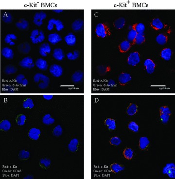
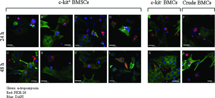
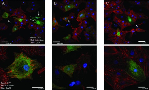
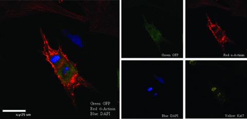
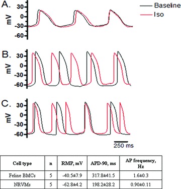
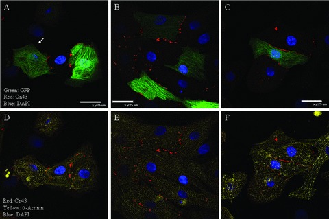
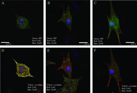
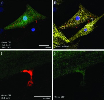
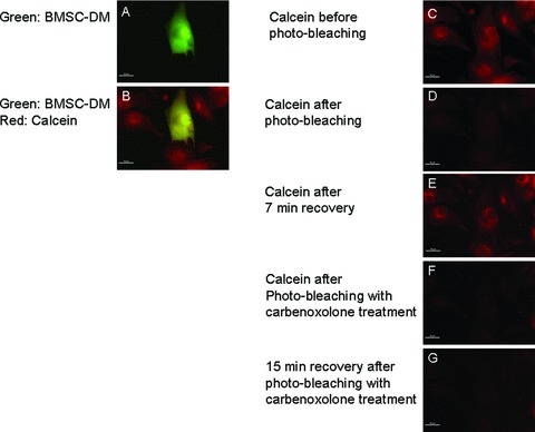
The utility of bone marrow cells (BMCs) to regenerate cardiac myocytes is controversial. The present study examined the capacity of different types of BMCs to generate functional cardiac myocytes. Isolated c-kit(+) BMCs (BMSCs), c-kit(+) and crude BMCs from the adult feline femur were membrane stained with PKH26 dye or infected with a control enhanced green fluorescence protein transcript (EGFP)-adenovirus prior to co-culture upon neonatal rat ventricular myocytes (NRVM). Co-cultured cells were immuno-stained for c-kit, alpha-tropomyosin, alpha-actinin, connexin 43 (Cx43) and Ki67 and analyzed with confocal microscopy. Electrophysiology of BMSC derived myocytes were compared to NRVMs within the same culture dish. Gap junction function was analyzed by fluorescence recovery after photo-bleaching (FRAP). BMCs proliferated and differentiated into cardiac myocytes during the first 48 hours of co-culturing. These newly formed cardiac myocytes were able to contract spontaneously or synchronously with neighboring NRVMs. The myogenic rate of c-kit(+) BMSCs was significantly greater than c-kit(+) and crude BMCs (41.2 +/- 2.1, 6.1 +/- 1.2, and 17.1 +/- 1.5%, respectively). The newly formed cardiac myocytes exhibited an immature electrophysiological phenotype until they became electrically coupled to NRVMs through functional gap junctions. BMSCs did not become functional myocytes in the absence of NRVMs. In conclusion, c-kit(+) BMSCs have the ability to transdifferentiate into functional cardiac myocytes.


Figures

Figure 1
Figure 1
Characterization of c‐kit+ bone marrow stem cells (BMSCs): C‐kit+ and c‐kit cells (A and C) were sorted using immuno‐magnetic beads from mononucleated bone marrow cells (BMCs). Our sorting method yielded c‐kit+ cells that were 1.6 ± 0.4% (n= 10) of the total BMCs. The isolated c‐kit+ BMSCs exhibited c‐kit immunofluorescence signals (C and D), while the c‐kit cells exhibited little if any c‐kit signals (A and B). Each cell type was detected to have low CD45 signals (B and D). The cardiac myocyte marker α‐Actinin was not detected in any of the cell types (A and C).
Figure 2
Figure 2
c‐Kit+‐BMSCs express cardiac contractile proteins when co‐cultured with NRVMs: c‐kit+ BMSCs, c‐kit, and crude BMCs were stained with a membrane dye PKH26 before being co‐cultured with NRVMs. The co‐culture was analyzed for the expression of cardiac contractile protein (α‐tropomyosin) at 24 h (A–F) and 48 h (G–L). During the 48 hours of co‐culturing, the c‐kit+ BMSCs expressed α‐tropomyosin. At 24 hours, the majority of the PKH26 stained BMSCs were small round shaped cells and only a few cells had α‐tropomyosin expression (A–D). Some cells were in mitosis, with (C) or without α‐tropomyosin expression (A). At 48 hours, the number of small round cells was decreased (H). The mitotic cells with α‐tropomyosin expression were still present (G) but the majority of the α‐tropomyosin expressing cells were larger (I and J). PKH26 stained c‐kit and crude BMCs were also found to express α‐tropomyosin at 48 hours (K and L); however, at 24 hours, α‐tropomyosin expression were rarely found in these cells (E and F). The percentage of c‐kit+, c‐kit, and crude BMCs with contractile protein expression at 24 hours was 15.4 ± 1.7, 4.0 ± 1.1, and 6.1 ± 1.2%, respectively. At 48 hours, these percentages increased to 41.1 ± 2.1, 12.3 ± 1.4, and 17.1 ± 1.5%, respectively. Red: PKH26; green: α‐tropomyosin; blue: DAPI.
Figure 3
Figure 3
c‐Kit+‐BMSCs differentiate into cardiac myocytes with organized sarcomeres: GFP expressing c‐kit+‐BMSCs were co‐cultured for 48 hours with NRVMs and analyzed for α‐actinin immunofluorescence. 38.6 ± 1.3%(n= 4) of the GFP expressing cells were observed to have organized α‐actinin (A, B, and C). White arrows indicate the cells magnified in the bottom panels. Dotted white arrows indicate GFP expressing BMSC derived non‐myocytes that are not expressing α‐actinin (A). Green: GFP; red: α‐Actinin; blue DAPI.
Figure 4
Figure 4
BMSC‐DMs are proliferative. Some GFP expressing BMSC‐DMs were found to be in the cell cycle.
Figure 5
Figure 5
BMSC‐DMs have electrophysiological properties of cardiac myocytes: The electrical properties of the BMSC‐DMs were related to the degree of coupling with NRVMs. BMSC‐DMs with minimal contact with NRVMs (A); BMSC‐DMs attached to a NRVM (B); NRVMs with minimal contact to other cells (C). Basal electrophysiological properties of BMSC‐DMs co‐cultured with NRVMs: BMSC‐DMs exhibited spontaneous action potentials (APs), whereas some NRVMs required electrical stimulation induced AP measurement. RMP: resting membrane potential; APD‐90, action potential duration at 90% repolarization.
Figure 6
Figure 6
Three representative examples of BMSC‐DMs that form gap junctions with neighboring cardiac myocytes: The GFP expressing BMSCs were co‐cultured with NRVMs. At 48 hours, the co‐culture was analyzed for α‐actinin and connexin 43 (C×43) immunofluorescence. GFP (green) expressing BMSC‐DMs localized C×43 (red) at the cell border with neighboring NRVMs (A, B, and C). Cardiac myocytes were identified by α‐actinin immunofluorescence (yellow: D, E, and F). C×43 was not detected between BMSC‐DMs and non‐myocytes (white arrow: A).
Figure 7
Figure 7
BMSC‐DMs in the cell cycle or those not making close connections with NRVMs did not have surface membrane C×43 localization; The BMSC‐DMs in the cell cycle (A and D) and the BMSC‐DMs having little (B and E) or no contact (C and F) with neighboring myocytes had punctuate, intracellular localized C×43. BMSC‐DMs with minimal contact with NRVMs had intense C×43 staining at the points of contact. Some BMSC‐DMs had small areas of contact with neighboring NRVMs (G and H) and C×43 immunofluorescence was intense at these areas (I and J).
Figure 7
Figure 7
BMSC‐DMs in the cell cycle or those not making close connections with NRVMs did not have surface membrane C×43 localization; The BMSC‐DMs in the cell cycle (A and D) and the BMSC‐DMs having little (B and E) or no contact (C and F) with neighboring myocytes had punctuate, intracellular localized C×43. BMSC‐DMs with minimal contact with NRVMs had intense C×43 staining at the points of contact. Some BMSC‐DMs had small areas of contact with neighboring NRVMs (G and H) and C×43 immunofluorescence was intense at these areas (I and J).
Figure 8
Figure 8
FRAP shows that BMSC‐DMs are coupled to neighboring cardiac myocytes: The GFP expressing BMSCs were co‐cultured with NRVMs. At hour 48, the co‐culture was loaded with calcein red‐orange‐AM (calcein) for 15 minutes (A, B, and C) and then photo‐bleached (D). The photobleached calcein signal recovered after 7 minutes (E). This recovery was not observed after treatment with the gap junction blocker carbenoxolone (100 μM) (G).

Similar articles

Cited by

References

    1. Kajstura J, Cheng W, Reiss K, Clark WA, Sonnenblick EH, Krajewski S, Reed JC, Olivetti G, Anversa P. Apoptotic and necrotic myocyte cell deaths are independent contributing variables of infarct size in rats. Lab Invest. 1996; 74(1): 86–107. - PubMed
    1. Shorofsky SR, Aggarwal R, Corretti M, Baffa JM, Strum JM, Al‐Seikhan BA, Kobayashi YM, Jones LR, Wier WG, Balke CW. Cellular mechanisms of altered contractility in the hypertrophied heart: big hearts, big sparks. Cir Res. 1999; 84(4): 424–434. - PubMed
    1. Bristow MR, Kantrowitz NE, Ginsburg R, Fowler MB. Beta‐adrenergic function in heart muscle disease and heart failure. J Mol Cell Cardiol. 1985; 17(Suppl 2): 41–52. - PubMed
    1. Shin J, Johnson JA. beta‐Blocker pharmacogenetics in heart failure. Heart Fail Rev. 2008 April 24. (Epub ahead of print) - PMC - PubMed
    1. Ramasubbu K, Mann DL, Deswal A. Anti‐angiotensin therapy: new perspectives. Cardiol Clin. 2007; 25(4): 573–580; vi–vii. - PubMed

Publication types

MeSH terms

Substances