. 2010 Jun;76(6):1340-57.
doi: 10.1111/j.1365-2958.2010.07181.x. Epub 2010 Apr 23.

Cathepsin L occupies a vacuolar compartment and is a protein maturase within the endo/exocytic system of Toxoplasma gondii

Affiliations

Cathepsin L occupies a vacuolar compartment and is a protein maturase within the endo/exocytic system of Toxoplasma gondii

Fabiola Parussini et al. Mol Microbiol. 2010 Jun.

Abstract

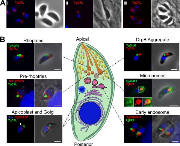
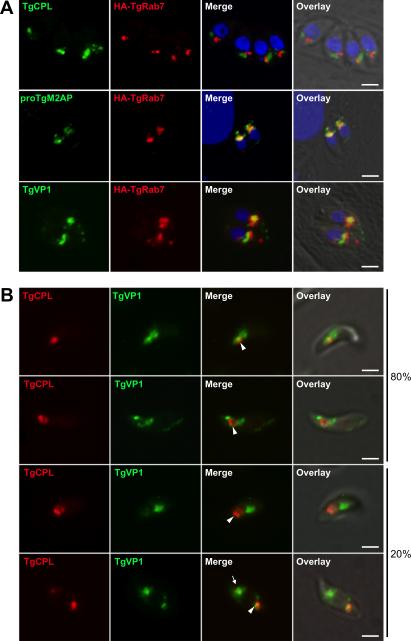
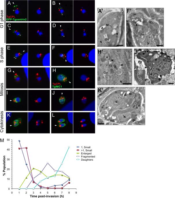
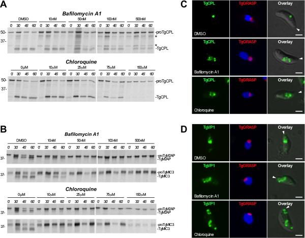
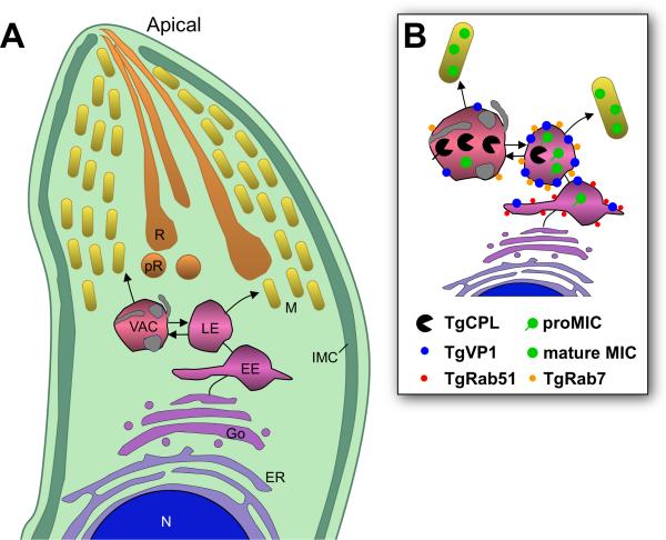
Regulated exocytosis allows the timely delivery of proteins and other macromolecules precisely when they are needed to fulfil their functions. The intracellular parasite Toxoplasma gondii has one of the most extensive regulated exocytic systems among all unicellular organisms, yet the basis of protein trafficking and proteolytic modification in this system is poorly understood. We demonstrate that a parasite cathepsin protease, TgCPL, occupies a newly recognized vacuolar compartment (VAC) that undergoes dynamic fragmentation during T. gondii replication. We also provide evidence that within the VAC or late endosome this protease mediates the proteolytic maturation of proproteins targeted to micronemes, regulated secretory organelles that deliver adhesive proteins to the parasite surface during cell invasion. Our findings suggest that processing of microneme precursors occurs within intermediate endocytic compartments within the exocytic system, indicating an extensive convergence of the endocytic and exocytic pathways in this human parasite.


Figures

Fig. 1
Fig. 1. TgCPL occupies a novel apical organelle
A. TgCPL localization in formaldehyde fixed RH parasites by immunofluorescence using MαTgCPL showing that TgCPL occupies a single apical localization in extracellular (i) and newly invaded parasites (ii). In contrast, TgCPL showed a punctate distribution in tachyzoites undergoing intracellular replication (iii). B. Dual staining of TgCPL with previously defined exocytic and endocytic markers. Parasites were stained with antibodies to TgROP2 (rhoptries), proTgROP4 (prerhoptry), TgGRASP-mRFP (Golgi cisternae), TgDrpB (cytoplasmic aggregate), TgAMA1 (micronemes), or TgRab51HA (EE). For the parasite illustration, the cytoplasm and pellicular membranes (plasma membrane and inner membrane complex) are shown in shades of green, DNA containing structures (nucleus and apicoplast) are blue, the early exocytic pathway (ER and Golgi) is shown in shades of purple, the endocytic system (EE, LE, and VAC) in shades of pink, and the late exocytic system in shades of orange-yellow. Scale bar, 2μm.
Fig. 2
Fig. 2. The VAC is juxtaposed to the LE, which contain proTgM2AP and TgVP1
A. RH parasites were transiently transfected with a plasmid expressing the LE marker TgRab7HA and intracellular parasites were fixed and processed for IFA 24 h post-transfection. B. Dual staining of TgCPL and TgVP1 in extracellular tachyzoites. TgCPL is concentrated in the VAC along with some TgVP1 (arrowhead) in most parasites. TgVP1 additionally occupies sites that are often adjacent to the VAC and in some cases also contain a minority population of TgCPL (arrow). Scale bar, 2 mm.
Fig. 3
Fig. 3. Ultrastructure of the VAC
Extracellular tachyzoites were immunolabeled with MαTgCPL antibodies before incubation with 10 nm protein A-gold particles. A. A globoid VAC surrounded by a single membrane containing TgCPL is observed in the apical area enriched in micronemes (Mi) and rhoptries (R). Magnifications (A’, A’’) reveal the presence of intraluminal vesicles with dense core material. B and inset. Internal vesicles delineated by a double membrane (arrow) are visible that may be the result of engulfment of an incoming vesicle by the VAC. C and C’. Deep invaginations of the VAC limiting membrane. D. Outward deformation of the multivesicular endosomal membrane. E. Tubulovesicular structures decorated by TgCPL may either result from the budding of the limiting membrane of the VAC or correspond to endosomal tubules fusing with the VAC. Scale bars are 200 nm, except 100 nm in inset in C.
Fig. 4
Fig. 4. Dynamic fragmentation of the VAC during intracellular replication
TgCPL distribution during intracellular replication was studied by staining EGFP-Centrin2 expressing parasites with RαTgCPL and MαGFP (to enhance the signal) (A-G) or RH parasites with MαTgCPL and RαTgIMC1 (H-L). Infected host cells were fixed at 1 h intervals 1-8 h post-invasion. The apical end of the parasite is indicated with an arrowhead and the centrosome is marked with an asterisk. A’, F’, H’, J’ and K’. Cryoimmunoelectron micrographs of MαTgCPL immunogold (10 nm) staining of intracellular tachyzoites in different stages of endodyogeny. Apicoplast, A; Golgi, Go; Rhoptry, R; Microneme, Mi; Nucleus, N, Parasitophorous Vacuole, PV; Residual Body, RB; VAC, black arrowhead; Apical tip of the parasites, white arrowhead. See text for descriptions. Scale bars, 200 nm. M. Quantification of TgCPL staining patterns during parasite replication. Data shown are the combined results of three independent experiments.
Fig. 5
Fig. 5. rTgCPL cleavage specificity is consistent with the propeptide cleavage site of several proMICs
A-B. Mapping P2 and P3 cleavage specificities of TgCPL and TgCPB. rTgCPL and rTgCPB (10 nM each) were screened against a Ala-P3-P2-Arg-ACC peptide substrate library (10μM) with all possible combinations of natural amino acids (except Cys) at the P2 and P3 positions (Salisbury et al., 2002; Gosalia et al., 2005). Initial rate velocities were measured by fluorescence in 384-well plates and are displayed as a heat map with color intensity indicating the rate of cleavage. Note that the strong preference of rTgCPB for P3 Pro might not be accurately indicated for peptides also having a highly preferred P2 residue (Phe, Leu, Val) because these reactions may have proceeded so quickly that they reached plateau phase before values could be measured. The experiment was performed three times with consistent results. C. Alignment of the r100proTgCPL autocleavage site with the propeptide cleavage sites of proMICs. Sequences were manually aligned (without gaps) with respect to the principal propeptide cleavage site (large arrow). A minor cleavage site of r100proTgCPL is also shown (small arrow). Identical residues within each group are shown in black boxes and similar residues are shown in grey boxes. Similar amino acids were classified as: non-polar (G, A, V, L, M, I); aromatic (F, Y, W); polar, uncharged (S, T, C, P, N, Q); polar, positively charged (K, R, H); or polar, negatively charged (D, E). TgMIC11 has an internal propeptide that is excised by two cleavage events at site 1 and site 2 (Harper et al., 2004). Protein domains are as follows: EGF, epidermal growth factor; Dom. I, domain I; Dom. II, domain II, Dom. III, domain III; CBL, chitin binding-like; TSR, thrombospondin repeat; Cys, cysteine protease. Transmembrane anchors are illustrated as black rectangles. Domain structures of TgMIC5 and TgMIC11 are unknown since they lack homology to other proteins. The P2 position of the proMIC propeptide cleavage sites is marked with an asterisk to indicate the highest level of conservation. Cleavage sites were defined in the following studies: TgM2AP (Rabenau et al., 2001), TgMIC6 (Opitz et al., 2002), TgAMA1 (Howell et al., 2005), TgMIC3 (Garcia-Réguet et al., 2000), TgSPATR (Kawase et al., 2007), r100proTgCPL (present study), TgMIC5 (Brydges et al., 2000), TgMIC11 (Harper et al., 2004).
Fig. 6
Fig. 6. TgCPL-deficient parasites show impaired maturation of proTgM2AP and proTgMIC3, and are defective in cell invasion
A. Pulse metabolically labeled RH or RHΔcpl parasites were either kept on ice (0 min) or chased with medium containing unlabelled Met/Cys for the indicated times. MIC proteins were immunoprecipitated and analyzed by SDS-PAGE and autoradiography. Arrows indicate positions of the immature (upper) and mature (lower) forms of each MIC protein. B-E. Quantification of MIC maturation in RH (squares) and RHΔcpl (circles) parasites is shown. Values are mean±s.d. of three (TgMIC6, TgAMA1), five, (TgMIC3), or six (TgM2AP) experiments and were calculated as described in figure legend 4. F. RHΔcpl parasites are less invasive than RH parasites. Parasites were allowed to invade HFF cells for 30 min prior to fixation. Extracellular parasites were stained with RαTgSAG1 before permeabilization and staining of intracellular parasites with a monoclonal antibody to TgSAG1.
Fig. 7
Fig. 7. rTgCPL correctly processes rproTgM2AP in a low pH dependent manner
A. Immunoblot probed with rabbit α-His tag showing a pH dependent TgM2AP propeptide cleavage mediated by rTgCPL. Affinity purified rproTgM2AP (400 ng) was incubated with rTgCPL (40 ng) at each pH value for the indicated times at 37°C. Note the low pH and time dependent production of a band that comigrates with rTgM2AP.
Fig. 8
Fig. 8. In vivo maturation of proTgCPL and proTgM2AP are differentially sensitive to pH antagonists
A. Representative autoradiographs showing inhibition of processing in parasite treated with pH-altering substances. RH parasites were pretreated for 15 min with the indicated concentrations of bafilomycin A1 or chloroquine before pulse labeling with [35S] Met/Cys for 10 min, chasing for the indicated times, and immunoprecipitation with Rαr100TgCPL. Asterisks indicate intermediate processed forms (48 kDa and 32 kDa) of proTgCPL. B. Same as A except immunoprecipitations were performed with RαrTgM2AP and mAb T4.2F3 against TgMIC3. Molecular weight markers are expressed in kDa. C. Bafilomycin A1 and chloroquine induced enlargement of the VAC. Representative immunofluorescence images showing TgCPL localization in RH parasites expressing TgGRASP-mRFP after treatment with pH antagonists. Parasites were treated with DMSO (upper panel); 500 nM bafilomycin A1 (middle panel); or 150 μM chloroquine (lower panel) for 15 min at RT, and then incubated for another 1h at 37°C. D. Bafilomycin A1 and chloroquine induced swelling and disassembling of the LE. Representative immunofluorescence images showing TgVP1 localization in RH parasites expressing TgGRASP-mRFP after treatment with DMSO (upper panel); 500 nM bafilomycin A1 (middle panel); or 150 μM chloroquine (lower panel) as described above. Arrowheads indicate the apical end of the parasite.
Fig. 9
Fig. 9. TgCPL and proTgM2AP encounter each other in microdomains along the endo/exocytic pathway
A. Dual staining of TgCPL and proTgM2AP during daughter cell formation was studied in fixed intracellular (G1, S, M) or extracellular (E) RH parasites by double immunolocalization using MαTgCPL and RαproTgM2APpep. TgCPL and proM2AP are located on adjacent dynamic compartments with small areas or microdomains where the overlap is evident at different stages of daughter cell formation. Arrows indicate the areas where small amounts of TgCPL have access to proTgM2AP. The inset is a higher magnification of the region indicated by dotted frames. B-E. Double cryoimmunoelectron microscopy of extracellular tachyzoites immunolabeled with anti-TgCPL (revealed with protein A-gold particles of 10 nm), and anti-proTgM2AP (C; 5 nm gold) or anti-TgM2AP. D, E, and F. (5 nm gold). Arrows pinpoint the co-distribution of TgCPL and proTgM2AP. Also note the concentration of micronemes (Mi) containing TgCPL and TgM2AP around the VAC (panels E and F). Scale bars are 100 nm.
Fig. 10
Fig. 10. Hypothetical model of trafficking and maturation in the endo/exocytic pathway
A. The model indicates that MIC protein complexes are synthesized in the ER before transiting through the Golgi apparatus and a series of post-Golgi endosomal compartments including the EE, the LE, and possibly the VAC. Rather than the TGN being the sorting station from where the budding of immature secretory granules primarily occurs as in higher eukaryotes, our model proposes that micronemes are formed by budding from an endocytic compartment, either the LE or the VAC. Dashed lines indicate possible alternative routing of proTgCPL from the ER or Golgi to the VAC. B. The model further proposes that proteolytic maturation of proMICs mediated by TgCPL and other proteases occurs within these compartments and possibly also in nascent micronemes. Acidification of the intralumenal environment of both compartments could be an important endogenous factor modulating the autoactivation of proTgCPL and limited proteolysis by TgCPL or alternative maturases.

Similar articles

Cited by

References

    1. Barrett AJ. Cathepsin G. Methods Enzymol. 1981;80(Pt C):561–565. - PubMed
    1. Binder EM, Lagal V, Kim K. The prodomain of Toxoplasma gondii GPI-anchored subtilase TgSUB1 mediates its targeting to micronemes. Traffic. 2008;9:1485–1496. - PMC - PubMed
    1. Botero-Kleiven S, V F., Lindh J, Richter-Dahlfors A, von Euler A, Wahlgren M. Receptor-mediated endocytosis in an apicomplexan parasite (Toxoplasma gondii). Exp Parasitol. 2001;98:134–44. - PubMed
    1. Bradley PJ, Boothroyd JC. The pro region of Toxoplasma ROP1 is a rhoptry-targeting signal. Int J Parasitol. 2001;31:1177–1186. - PubMed
    1. Breinich MS, Ferguson DJ, Foth BJ, van Dooren GG, Lebrun M, Quon DV, et al. A dynamin is required for the biogenesis of secretory organelles in Toxoplasma gondii. Curr Biol. 2009;19:277–286. - PMC - PubMed

Publication types