

Duodenal gangliocytic paraganglioma showing lymph node metastasis: a rare case report
- PMID: 20444291
- PMCID: PMC2874790
- DOI: 10.1186/1746-1596-5-27
Duodenal gangliocytic paraganglioma showing lymph node metastasis: a rare case report
Abstract
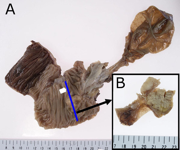
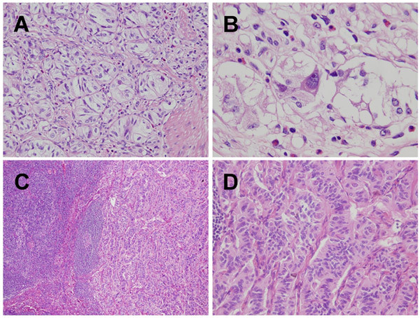
We describe a case of duodenal gangliocytic paraganglioma showing lymph node metastasis. A 61-year-old Japanese man underwent pylorus preserving pancreaticoduodenectomy to remove a tumor at the papilla of Vater. The section of the tumor extending from the mucosa to submucosa of the duodenum was sharply demarcated, solid, and white-yellowish. Neither necrosis nor hemorrhage was present. Histological examination confirmed the immunohistochemical identification of three components comprising epithelioid cells, spindle-shaped cells, and ganglion-like cells. Epithelioid cells showed positive reactivity for synaptophysin, somatostatin, and CD56. In contrast, spindle-shaped cells showed positive reactivity for S-100 protein, but not for synaptophysin, somatostatin or CD56. Furthermore, we found lymph node metastasis despite lack of bcl-2 and p53 expression. In addition to the rarity of the tumor, we are describing here the present case suggests the malignant potency of the tumor despite lack of acceptable prognostic indicators for neuroendocrine tumor.
Figures


Similar articles
-
A gangliocytic patially glandular paraganglioma with lymph node metastasis.Diagn Pathol. 2014 Mar 20;9:63. doi: 10.1186/1746-1596-9-63. Diagn Pathol. 2014. PMID: 24649939 Free PMC article.
-
Gangliocytic paraganglioma: case report and review of the literature.J Gastrointest Surg. 2007 Oct;11(10):1351-4. doi: 10.1007/s11605-007-0217-9. Epub 2007 Jul 25. J Gastrointest Surg. 2007. PMID: 17653595 Review.
-
Malignant gangliocytic paraganglioma of the duodenum with distant metastases and a lethal course.World J Gastroenterol. 2014 Nov 7;20(41):15454-61. doi: 10.3748/wjg.v20.i41.15454. World J Gastroenterol. 2014. PMID: 25386095 Free PMC article. Review.
-
Duodenal gangliocytic paraganglioma: report of two cases and review of literature.Int J Clin Exp Pathol. 2015 Sep 1;8(9):9752-9. eCollection 2015. Int J Clin Exp Pathol. 2015. PMID: 26617685 Free PMC article. Review.
-
An unusual case of duodenal obstruction-gangliocytic paraganglioma.J Hepatobiliary Pancreat Surg. 2009;16(4):562-5. doi: 10.1007/s00534-009-0092-8. Epub 2009 Jun 11. J Hepatobiliary Pancreat Surg. 2009. PMID: 19517054
Cited by
-
Duodenal gangliocytic paraganglioma: A very rare cause for upper gastrointestinal bleeding: Case report with review of literature.Int J Surg Case Rep. 2020;75:408-412. doi: 10.1016/j.ijscr.2020.09.129. Epub 2020 Sep 23. Int J Surg Case Rep. 2020. PMID: 33002850 Free PMC article.
-
Endoscopic resection of gangliocytic paraganglioma of the duodenum: a case report.Clin J Gastroenterol. 2020 Apr;13(2):203-208. doi: 10.1007/s12328-019-01043-0. Epub 2019 Sep 18. Clin J Gastroenterol. 2020. PMID: 31535284
-
Literature survey on epidemiology and pathology of gangliocytic paraganglioma.BMC Cancer. 2011 May 20;11:187. doi: 10.1186/1471-2407-11-187. BMC Cancer. 2011. PMID: 21599949 Free PMC article.
-
A gangliocytic patially glandular paraganglioma with lymph node metastasis.Diagn Pathol. 2014 Mar 20;9:63. doi: 10.1186/1746-1596-9-63. Diagn Pathol. 2014. PMID: 24649939 Free PMC article.
-
Bronchogenic Gangliocytic Paraganglioma.J Bronchology Interv Pulmonol. 2020 Jul;27(3):212-215. doi: 10.1097/LBR.0000000000000667. J Bronchology Interv Pulmonol. 2020. PMID: 32205710 Free PMC article. No abstract available.
References
-
- Sundararajan V, Robinson-Smith TM, Lowy AM. Duodenal gangliocytic paraganglioma showing lymph node metastasis: a case report and review of the literature. Arch Pathol Lab Med. 2003;127:139–141. - PubMed
-
- de Krijger RR, Harst E van der, Ham F van der, Stijnen T, Dinjens WN, Koper JW, Bruining HA, Lamberts SW, Bosman FT. Prognostic value of p53, bcl-2, and c-erbB-2 protein expression in phaeochromocytomas. J Pathol. 1999;188:51–55. doi: 10.1002/(SICI)1096-9896(199905)188:1<51::AID-PATH310>3.0.CO;2-R. - DOI - PubMed
-
- Cadden IS, Atkinson AB, Johnston BT, Pogue K, Connolly R, McCance D, Ardill JE, Russell CF, McGinty A. Cyclooxygenase-2 expression correlates with phaeochromocytoma malignancy: evidence for a Bcl-2-dependent mechanism. Histopathology. 2007;51:743–751. doi: 10.1111/j.1365-2559.2007.02846.x. - DOI - PubMed
Publication types
MeSH terms
Substances
LinkOut - more resources
Full Text Sources
Medical
Research Materials
Miscellaneous
