. 2010 May 5;11(5):379-89.
doi: 10.1016/j.cmet.2010.03.013.

Loss of insulin signaling in vascular endothelial cells accelerates atherosclerosis in apolipoprotein E null mice

Affiliations

Loss of insulin signaling in vascular endothelial cells accelerates atherosclerosis in apolipoprotein E null mice

Christian Rask-Madsen et al. Cell Metab. .

Abstract

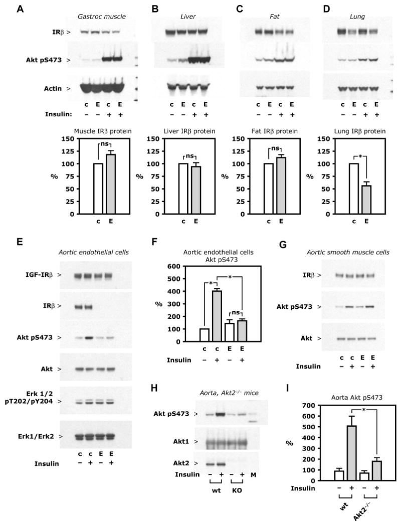
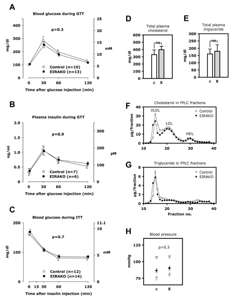
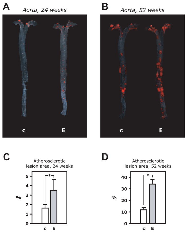
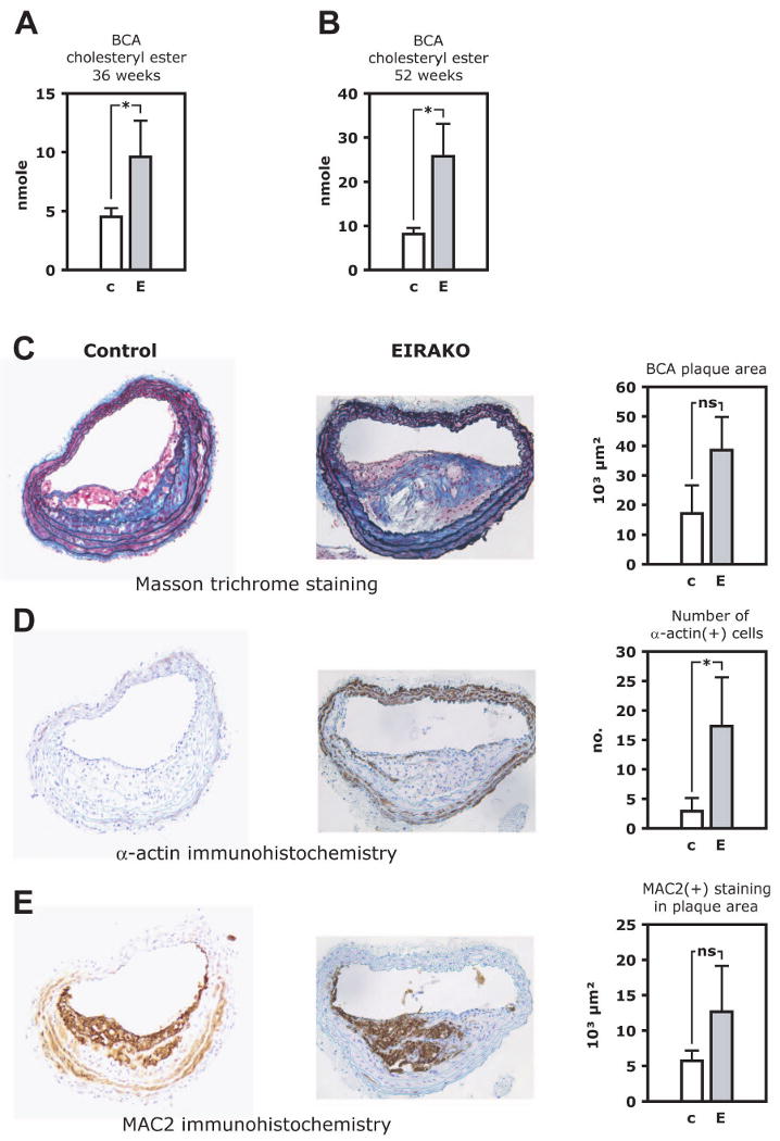
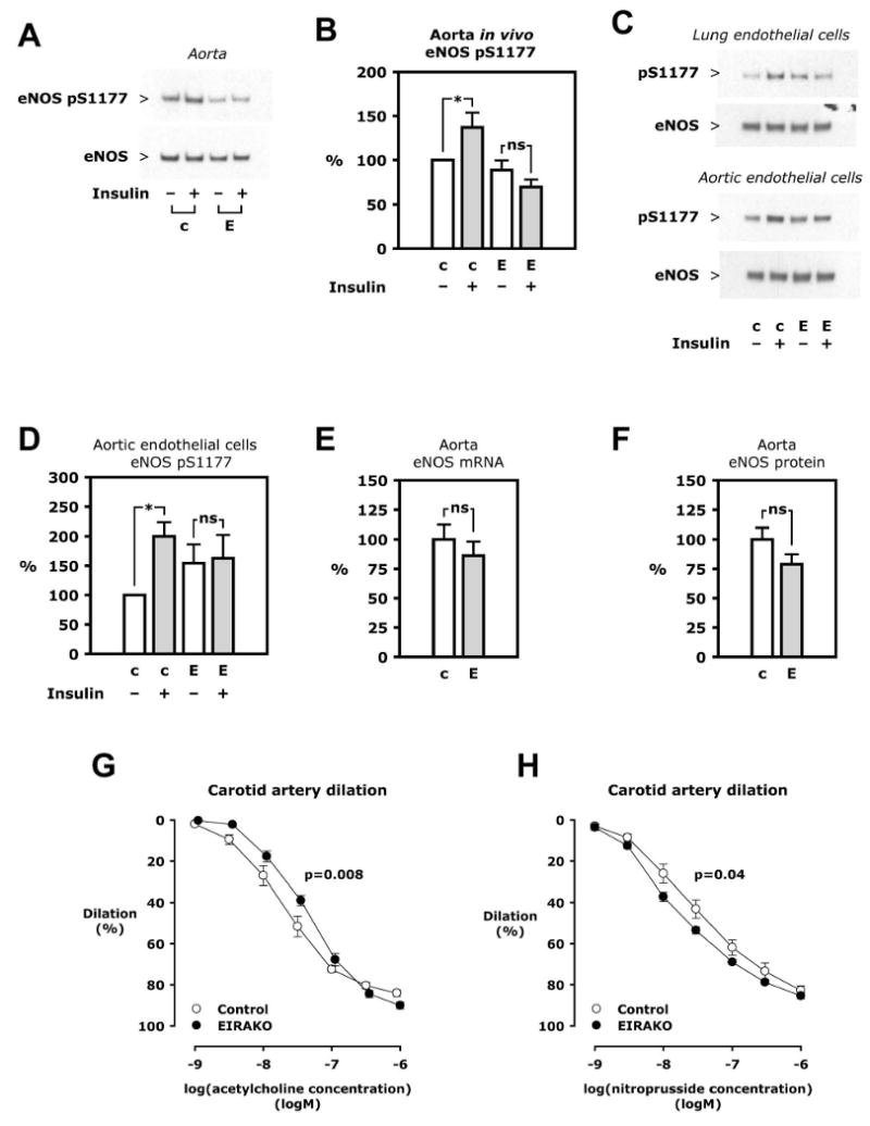
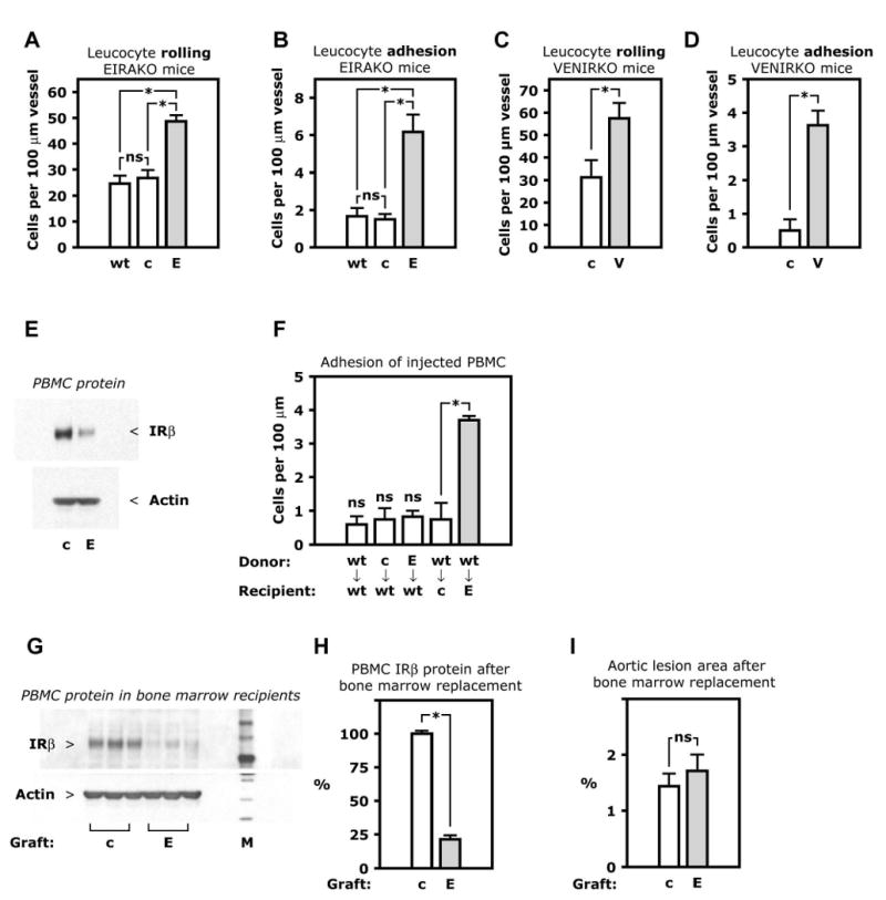
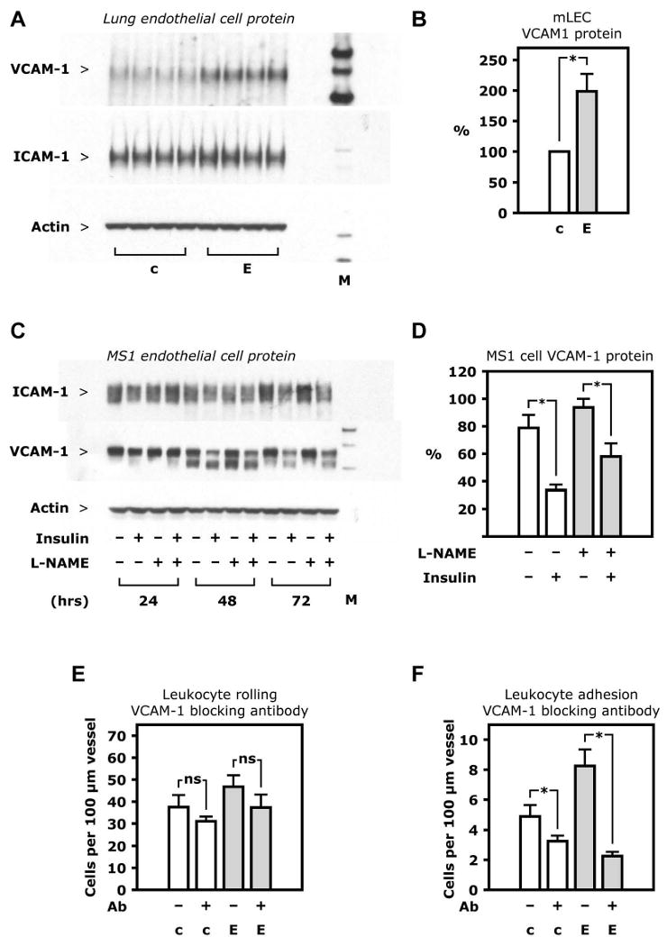
To determine whether insulin action on endothelial cells promotes or protects against atherosclerosis, we generated apolipoprotein E null mice in which the insulin receptor gene was intact or conditionally deleted in vascular endothelial cells. Insulin sensitivity, glucose tolerance, plasma lipids, and blood pressure were not different between the two groups, but atherosclerotic lesion size was more than 2-fold higher in mice lacking endothelial insulin signaling. Endothelium-dependent vasodilation was impaired and endothelial cell VCAM-1 expression was increased in these animals. Adhesion of mononuclear cells to endothelium in vivo was increased 4-fold compared with controls but reduced to below control values by a VCAM-1-blocking antibody. These results provide definitive evidence that loss of insulin signaling in endothelium, in the absence of competing systemic risk factors, accelerates atherosclerosis. Therefore, improving insulin sensitivity in the endothelium of patients with insulin resistance or type 2 diabetes may prevent cardiovascular complications.


Figures

Figure 1
Figure 1. Insulin receptor expression and insulin signaling
A-D. Insulin 5 mU/g i. v. was given to mice fasted overnight and tissues collected after 5 minutes. Representative Western blots (top) show insulin receptor-β (IRβ) protein expression and insulin-stimulated Akt Ser473 phosphorylation in lysate of the tissues indicated; also shown are mean values of IRβ protein after densitometry of Western blots from 3 independent experiments (bottom). E-F. Aorta was digested by collagenase and endothelial cells were isolated by immunoselection with magnetic microbeads complexed with ICAM2 antibody, with aortic smooth muscle cells grown from the ICAM2-negative fraction. Cultures were treated with insulin (10 nM, 5 minutes) after 24 hours of serum starvation. E. Representative Western blots from whole cell lysate of primary culture of aortic endothelial cells are shown. F. Mean values of Akt Ser473 phosphorylation was based on densitometry of Western blots from 3 independent experiments. G. Western blots from whole cell lysate of aortic smooth muscle cells representative of 3 independent experiments. H-I. Insulin 10 mU/g was given i. v. in wild-type and Akt2−/− mice. After 5 minutes, the aorta was isolated and snap frozen. H. Representative Western blots of aorta lysate. I. Mean values of Akt Ser473 phosphorylation based on data from 10 wild-type and 13 Akt2−/− mice. See also Figure S1. Abbreviations: c, control mice; E, EIRAKO mice; wt, wild-type mice.
Figure 2
Figure 2. Whole-body glucose tolerance and insulin sensitivity, plasma lipids, blood pressure
A. Blood glucose during glucose tolerance test. B. Plasma insulin during glucose tolerance test. C. Plasma glucose during insulin tolerance test. D-G. Plasma lipids were measured in 12 EIRAKO mice and 11 littermate controls after a 4-6 hours fast. Plasma fractions were obtained by fast protein liquid chromatography (FPLC) in a subset of 3 EIRAKO mice and 3 littermate controls. D. Total cholesterol in plasma. E. Total triglycerides in plasma. F. Cholesterol concentration in FPLC fractions of plasma. G. Triglyceride concentration in FPLC fractions of plasma. H. Blood pressure was measured by tail vein plethysmography in 14 EIRAKO mice and 12 littermate controls. Mean values for systolic and diastolic blood pressure (open triangles) as well as mean blood pressure (closed circles) are shown.
Figure 3
Figure 3. Atherosclerotic lesion size in the aorta
A. Microphotographs of aortas from an EIRAKO mouse and its littermate control at 24 weeks of age in the en face flat preparation after staining with Sudan IV. B. Summary data from quantitation of atherosclerotic lesion area relative to total area of the aorta in 10 EIRAKO mice and 8 littermate controls at 24 weeks of age. C. Stained aortas at 52 weeks of age. D. Summary data from 9 EIRAKO animals and 11 littermate controls at 52 weeks of age
Figure 4
Figure 4. Brachiocephalic artery lesion size and atherosclerotic plaques histology
Lipids were quantitatively extracted from the brachiocephalic artery, which was then paraffin embedded and cross-sectioned. Cholesteryl ester was measured in lipid extracts by mass spectrometry. A. Cholesteryl ester content in the brachiocephalic artery at 36 weeks. B. Colesteryl ester content in the brachiocephalic artery at 52 weeks. C-E. Histological staining and immunohistochemistry of brachiocephalic artery cross-sections. Representative images are shown (left), as are mean values of quantitative analysis (right). C. Masson trichrome stain, with collagen staining blue. D. MAC-2 immunohistochemistry to visualize plaque area occupied by macrophages. E. α-smooth muscle cell actin immunohistochemistry to identify vascular smooth muscle cells. See also Figure S2.
Figure 5
Figure 5. eNOS regulation
A. Insulin 5 mU/g was injected i. v. and the aorta removed after 5 minutes. Western blotting was performed on aorta lysate. B. Mean values of eNOS Ser1177 phosphorylation relative to eNOS based on densitometry of Western blots of aorta from 6 sets of 4 animals. C. Primary cultures of lung and aortic endothelial cells isolated from an EIRAKO mouse and its littermate control were treated with insulin (100 nM in lung endothelial cells, 10 nM in aortic endothelial cells, 5 minutes). Representative Western blots are shown. D. Mean values of eNOS Ser1177 phosphorylation relative to eNOS expression in aortic endothelial cells from 3 independent experiments. E. eNOS mRNA expression in aorta measured by real-time PCR, mean results from 11 EIRAKO mice and 9 controls. F. eNOS protein in aorta based on densitometry of Western blots and normalized to actin, mean results from 11 EIRAKO mice and 8 controls. G-H. The carotid artery was isolated, mounted, and pressurized in a myograph. Graphs show mean vasodilation using arteries from 6 control and 6 EIRAKO mice. G. Concentration-response study using acetylcholine. H. Concentration-response study using sodium nitroprusside.
Figure 6
Figure 6. Leukocyte-endothelial cell interaction in vivo
A-D. Circulating leukocytes were fluorescently labeled by i. v. injection of rhodamine and visualized in peri-intestinal, post-capillary venules by intravital microscopy. Mean numbers of rolling or firmly adhering leukocytes in 3-4 animals are shown. A. Leukocyte rolling in EIRAKO mice and controls. B. Leukocyte adhesion in EIRAKO mice. C. Leukocyte rolling in VENIRKO mice and controls, both wild-type for apoE. D. Leukocyte adhesion in VENIRKO mice and controls. E. Western blotting of lysate of peripheral blood mononuclear cells (PBMC). F. Adhesion in vivo of transferred mononuclear cells. PBMC were freshly isolated from one donor animal, labeled with rhodamine ex vivo, and injected i. v. in a single recipient animal. Mean numbers of firmly adhering PBMC, with genotypes of donor and recipient animals indicated, are shown (n=3-5). G-I. Bone marrow transplantation in apoE knockout mice, which were lethally irradiated and recieved a graft of bone marrow cells from EIRAKO or control mice. An atherogenic “Western” diet was started the day after transplantation. G. Western blotting of PBMC lysate 8 weeks after transplantation. H. Mean values for insulin receptor-β protein based on densitometry of Western blots of PBMC from 3 mice in each group of apoE knockout mice with EIRAKO or control bone marrow replacement. See also Figure S3. I. Mean values for atherosclerotic lesion area in the aorta in 11 recipients of EIRAKO mice bone marrow and 11 recipients of control bone marrow 8 weeks after bone marrow transplantation. Abbreviations: E, endothelial insulin receptor and apoE knockout (EIRAKO) mice; V, vascular endothelial receptor knockout (VENIRKO) mice; PBMC, peripheral blood mononuclear cells.
Figure 7
Figure 7. VCAM-1 regulation
A. Western blotting of whole cell lysate from lung endothelial cells isolated from one EIRAKO mouse and one control, with each sample loaded as 4 replicates. B. Mean values for VCAM-1 protein based on densitometry of Western blots of lung endothelial cell lysate from 4 pairs of EIRAKO mice and controls. C. Western blotting of whole cell lysate from MS1 endothelial cells. D. Mean values for ICAM-1 and VCAM-1 protein based on densitometry of Western blots from 3 independent experiments in MS1 endothelial cells. E-F. Leukocyte rolling on and firm adhesion to endothelium of peri-intestinal, post-capillary venules visualized by intravital microscopy after fluorescent labeling by i. v. injection of rhodamine. Measurements were repeated 60 minutes after i. v. injection of a VCAM-1 blocking antibody. Mean values from 4 pairs of animals 20-23 weeks of age are shown. E. Leukocyte rolling before and after VCAM-1 injection F. Leukocyte adhesion before and after VCAM-1 injection.

Similar articles

Cited by

References

    1. Abid MR, Shih SC, Otu HH, Spokes KC, Okada Y, Curiel DT, Minami T, Aird WC. A novel class of vascular endothelial growth factor-responsive genes that require forkhead activity for expression. J Biol Chem. 2006;281:35544–35553. - PubMed
    1. Aljada A, Saadeh R, Assian E, Ghanim H, Dandona P. Insulin inhibits the expression of intercellular adhesion molecule-1 by human aortic endothelial cells through stimulation of nitric oxide. J Clin Endocrinol Metab. 2000;85:2572–2575. - PubMed
    1. Atochin DN, Wang A, Liu VW, Critchlow JD, Dantas AP, Looft-Wilson R, Murata T, Salomone S, Shin HK, Ayata C, Moskowitz MA, Michel T, Sessa WC, Huang PL. The phosphorylation state of eNOS modulates vascular reactivity and outcome of cerebral ischemia in vivo. J Clin Invest. 2007;117:1961–1967. - PMC - PubMed
    1. Baumgartl J, Baudler S, Scherner M, Babaev V, Makowski L, Suttles J, McDuffie M, Tobe K, Kadowaki T, Fazio S, Kahn CR, Hotamisligil GS, Krone W, Linton M, Bruning JC. Myeloid lineage cell-restricted insulin resistance protects apolipoproteinE-deficient mice against atherosclerosis. Cell Metab. 2006;3:247–256. - PMC - PubMed
    1. Booth G, Stalker TJ, Lefer AM, Scalia R. Elevated ambient glucose induces acute inflammatory events in the microvasculature: effects of insulin. Am J Physiol Endocrinol Metab. 2001;280:E848–856. - PubMed

Publication types