. 2010 Jul 20;403(1):26-36.
doi: 10.1016/j.virol.2010.03.027. Epub 2010 May 4.

The structure of adeno-associated virus serotype 3B (AAV-3B): insights into receptor binding and immune evasion

Affiliations

The structure of adeno-associated virus serotype 3B (AAV-3B): insights into receptor binding and immune evasion

Thomas F Lerch et al. Virology. .

Abstract

Adeno-associated viruses (AAVs) are leading candidate vectors for human gene therapy. AAV serotypes have broad cellular tropism and use a variety of cellular receptors. AAV serotype 3 binds to heparan sulfate proteoglycan prior to cell entry and is serologically distinct from other serotypes. The capsid features that distinguish AAV-3B from other serotypes are poorly understood. The structure of AAV-3B has been determined to 2.6A resolution from twinned crystals of an infectious virus. The most distinctive structural features are located in regions implicated in receptor and antibody binding, providing insights into the cell entry mechanisms and antigenic nature of AAVs. We show that AAV-3B has a lower affinity for heparin than AAV-2, which can be rationalized by the distinct features of the AAV-3B capsid. The structure of AAV-3B provides an additional foundation for the future engineering of improved gene therapy vectors with modified receptor binding or antigenic characteristics.


Figures

Figure 1
Figure 1
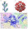
The structure of AAV-3B. (A) A portion of the 2mFo-DFc electron density surrounding part of the AAV-3B model shows the quality of the map calculated at 2.6Å resolution. (B) The jellyroll barrel subunit architecture of AAV-3B is conserved throughout the Parvovirus family. A cartoon representation of a single subunit is shown, surrounded by the transparent surfaces of neighboring subunits of the assembled capsid. Arrows mark the locations of the 5-, 3-, and 2-fold symmetry axes and are labeled. Loops are named after the core β-strands that they connect. The DE loop lines the interior of the 5-fold pore and forms the cylindrical protrusion surrounding the pore of the intact capsid (also shown in C). The HI loop covers part of the surface on the adjacent subunit (Chain E), lining the floor of a depression surrounding the cylinder. Two sections of each GH loop and two loops from neighboring subunit C comprise the prominent spikes that surround each 3-fold symmetry axis of the capsid. (C) Sixty AAV-3B subunits assemble to form the icosahedrally symmetric virus capsid. The viral asymmetric unit is bounded by 5-fold (pentagon), 3-fold (triangle), and 2-fold (oval) axes. The features on the capsid surface are formed by the subunit loops, shown in panel B.
Figure 2
Figure 2
Nucleotide binding inside the AAV-3B capsid, near the 3-fold symmetry axis. A single dAMP nucleotide was modeled in a pocket at the interior of the AAV-3B capsid The nucleotide is a fragment of the genomic single-stranded DNA that, through interactions with the capsid protein, adopts the capsid’s icosahedral symmetry. Thus, unlike most of the genomic DNA, its density remains strong through the symmetry averaging applied during the structure determination. The interior surface of one capsid subunit is shown in light pink, and the surfaces of 3-fold symmetric subunits are shown in light blue and light green. The dark grey mesh shows the 2.6Å mFo-DFc difference map (within 1.5Å of dAMP atoms), calculated with nucleotide coordinates omitted from the phasing model and contoured at 3.3 σ. The light grey mesh shows a 2mFo-DFc electron density map calculated using the 2.6Å data contoured at 1σ. Residues near the dAMP binding site are shown as sticks and are labeled. Density was also observed in an averaged map calculated using the untwinned 3.0Å data (not shown).
Figure 3
Figure 3
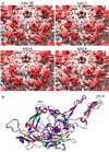
Comparing AAV serotype structures. (A) Molecular surfaces calculated from the atomic structures of AAV-3B, -2, -4, and -8. The triangular icosahedral asymmetric unit is outlined and is bounded by a 5-fold symmetry axis (pentagon), two 3-fold axes (triangles), and a 2-fold axis (oval). The most distinctive surface features correspond to the greatest differences in the subunit loop structures at “VR-I” (dashed circle) and “VR-IV” (solid circle) as illustrated in panel B. (B) Capsid subunits are overlaid with the secondary structure matching (SSM) algorithm (Krissinel and Henrick, 2004): AAV-3B (blue), -2 (Xie et al., 2002; red), -4 (Govindasamy et al., 2006; purple), and -8 (Nam et al., 2007; green). The greatest differences are in two of the variable regions (VR), I and IV.
Figure 4
Figure 4
Structural insights into receptor binding. Three of the sixty spikes on AAV-2 (A and C) and AAV-3B (B and D) are rendered as molecular surfaces. (A) Residues that have been shown previously to contact heparin directly (blue) or indirectly (teal) on AAV-2 are colored (O'Donnell, Taylor, and Chapman, 2009). Residues within the heparin binding footprint that are not conserved in AAV-3B are shaded lighter than conserved residues. (B) Residues from AAV-3B that align with the heparin contact residues in AAV-2 are similarly colored and shaded. The loss of the positive charge of R585 & R588 of AAV-2 might be partly compensated by the presence of R594 and/or R447 of AAV-3B. The electrostatic potential of AAV-2 (C) and AAV-3B (D) mapped onto the respective capsid surfaces is shown. Difference electron density for heparin bound to AAV-2, contoured at 7.5 σ (~5 estimated error units), is superimposed (O'Donnell, Taylor, and Chapman, 2009). The strongest density for the sulfonated heparin (negatively charged) was observed immediately adjacent to R585 and R588 of AAV-2 (C). (D) The corresponding region on the AAV-3B spike is less electro-positive. Two distinct basic patches on AAV-3B, one near R594 and the other near residue R447, are farther from the strongest heparin density seen in AAV-2, but are contained within (R594) or adjacent to (R447) the heparin binding footprint. The positive region that contains R447 is located on the opposite side of the spike relative to R585/R588 on AAV-2, and is most clearly seen here on the side of the 3-fold symmetric spike 3.
Figure 5
Figure 5
Specificity of MAb C37-B binding. A close-up view of a single 3-fold proximal spike on AAV-3B (A and C) and on AAV-2 (B and D) shows differences in surface topology and charge close to the mimotope of C37-B (AAV-2 residues 492–501, shown as sticks). C37-B blocks receptor binding to AAV-2, but does not bind AAV-3 (Wobus et al., 2000). Cartoon representations of two subunits (grey and yellow) are shown inside of a semi-transparent surface, colored by electrostatic potential. The mimotope is sandwiched between VR-IV and VR-VIII (protruding GH loops, labeled) from a neighboring subunit. The dimensions of a typical antibody footprint are drawn as a 30Å diameter circle, centered at N497 (AAV-3B) and N496 (AAV-2) in panels E and F, illustrating that C37-B would likely cover part of VR-IV and VR-VIII, including R585/R588 on AAV-2, perhaps explaining how C37-B blocks receptor binding to this serotype.

References

    1. Adams PD, Grosse-Kunstleve RW, Hung LW, Ioerger TR, McCoy AJ, Moriarty NW, Read RJ, Sacchettini JC, Sauter NK, Terwilliger TC. PHENIX: building new software for automated crystallographic structure determination. Acta Crystallogr D Biol Crystallogr. 2002;58(Pt 11):1948–1954. - PubMed
    1. Agbandje-McKenna M, Llamas-Saiz AL, Wang F, Tattersall P, Rossmann MG. Functional implications of the structure of the murine parvovirus, minute virus of mice. Structure. 1998;6(11):1369–1381. - PubMed
    1. Akache B, Grimm D, Pandey K, Yant SR, Xu H, Kay MA. The 37/67-kilodalton laminin receptor is a receptor for adeno-associated virus serotypes 8, 2, 3, and 9. J Virol. 2006;80(19):9831–9836. - PMC - PubMed
    1. Asokan A, Conway JC, Phillips JL, Li C, Hegge J, Sinnott R, Yadav S, DiPrimio N, Nam HJ, Agbandje-McKenna M, McPhee S, Wolff J, Samulski RJ. Reengineering a receptor footprint of adeno-associated virus enables selective and systemic gene transfer to muscle. Nat Biotechnol. 2010;28(1):79–82. - PMC - PubMed
    1. Asokan A, Hamra JB, Govindasamy L, Agbandje-McKenna M, Samulski RJ. Adeno-associated virus type 2 contains an integrin alpha5beta1 binding domain essential for viral cell entry. J Virol. 2006;80(18):8961–8969. - PMC - PubMed

Publication types

Substances