

The histone shuffle: histone chaperones in an energetic dance
- PMID: 20444609
- PMCID: PMC4004086
- DOI: 10.1016/j.tibs.2010.04.001
The histone shuffle: histone chaperones in an energetic dance
Abstract
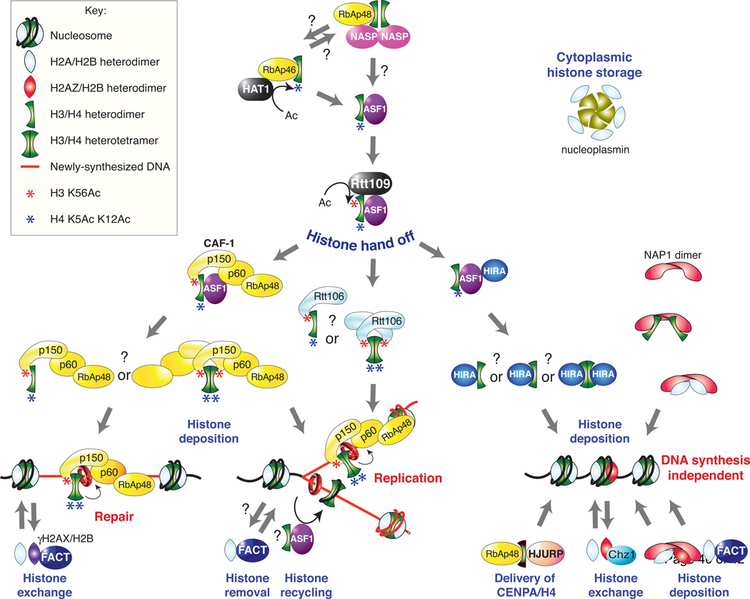
Our genetic information is tightly packaged into a rather ingenious nucleoprotein complex called chromatin in a manner that enables it to be rapidly accessed during genomic processes. Formation of the nucleosome, which is the fundamental unit of chromatin, occurs via a stepwise process that is reversed to enable the disassembly of nucleosomes. Histone chaperone proteins have prominent roles in facilitating these processes as well as in replacing old histones with new canonical histones or histone variants during the process of histone exchange. Recent structural, biophysical and biochemical studies have begun to shed light on the molecular mechanisms whereby histone chaperones promote chromatin assembly, disassembly and histone exchange to facilitate DNA replication, repair and transcription.
Copyright (c) 2010 Elsevier Ltd. All rights reserved.
Figures


Similar articles
-
Fly Fishing for Histones: Catch and Release by Histone Chaperone Intrinsically Disordered Regions and Acidic Stretches.J Mol Biol. 2017 Aug 4;429(16):2401-2426. doi: 10.1016/j.jmb.2017.06.005. Epub 2017 Jun 10. J Mol Biol. 2017. PMID: 28610839 Free PMC article. Review.
-
Interplay between histone variants and chaperones in plants.Curr Opin Plant Biol. 2024 Aug;80:102551. doi: 10.1016/j.pbi.2024.102551. Epub 2024 May 21. Curr Opin Plant Biol. 2024. PMID: 38776573 Review.
-
Methods to study histone chaperone function in nucleosome assembly and chromatin transcription.Methods Mol Biol. 2015;1288:375-94. doi: 10.1007/978-1-4939-2474-5_22. Methods Mol Biol. 2015. PMID: 25827892
-
Structure and function of histone chaperones in replication-coupled chromatin assembly.Curr Opin Struct Biol. 2025 Jun;92:103059. doi: 10.1016/j.sbi.2025.103059. Epub 2025 May 7. Curr Opin Struct Biol. 2025. PMID: 40339328 Review.
-
Mechanistic and structural insights into histone H2A-H2B chaperone in chromatin regulation.Biochem J. 2020 Sep 18;477(17):3367-3386. doi: 10.1042/BCJ20190852. Biochem J. 2020. PMID: 32941645 Review.
Cited by
-
Analysis of the Relationships between DNA Double-Strand Breaks, Synaptonemal Complex and Crossovers Using the Atfas1-4 Mutant.PLoS Genet. 2015 Jul 6;11(7):e1005301. doi: 10.1371/journal.pgen.1005301. eCollection 2015 Jul. PLoS Genet. 2015. PMID: 26147458 Free PMC article.
-
Analysis of histone chaperone antisilencing function 1 interactions.Methods Enzymol. 2012;512:223-41. doi: 10.1016/B978-0-12-391940-3.00010-X. Methods Enzymol. 2012. PMID: 22910209 Free PMC article.
-
Pyruvate Facilitates FACT-Mediated γH2AX Loading to Chromatin and Promotes the Radiation Resistance of Glioblastoma.Adv Sci (Weinh). 2022 Mar;9(8):e2104055. doi: 10.1002/advs.202104055. Epub 2022 Jan 20. Adv Sci (Weinh). 2022. PMID: 35048565 Free PMC article.
-
Histone H2A/H2B dimer exchange by ATP-dependent chromatin remodeling activities.Mol Cell. 2003 Dec;12(6):1599-606. doi: 10.1016/s1097-2765(03)00499-4. Mol Cell. 2003. PMID: 14690611 Free PMC article.
-
Unzipping single DNA molecules to study nucleosome structure and dynamics.Methods Enzymol. 2012;513:29-58. doi: 10.1016/B978-0-12-391938-0.00002-1. Methods Enzymol. 2012. PMID: 22929764 Free PMC article.
References
-
- Olins AL, Olins DE. Spheroid chromatin units (v bodies) Science. 1974;183:330–332. - PubMed
-
- Kornberg RD. Chromatin structure: a repeating unit of histones and DNA. Science. 1974;184:868–871. - PubMed
-
- Kornberg RD. Structure of chromatin. Annu Rev Biochem. 1977;46:931–954. - PubMed
-
- Luger K, et al. Crystal structure of the nucleosome core particle at 2.8 A resolution. Nature. 1997;389:251–260. - PubMed
-
- Van Holde KE. Chromatin. Springer-Verlag; 1989.
Publication types
MeSH terms
Substances
Grants and funding
LinkOut - more resources
Full Text Sources
Miscellaneous
