

Interconversion of the specificities of human lysosomal enzymes associated with Fabry and Schindler diseases
- PMID: 20444686
- PMCID: PMC2898384
- DOI: 10.1074/jbc.M110.118588
Interconversion of the specificities of human lysosomal enzymes associated with Fabry and Schindler diseases
Abstract
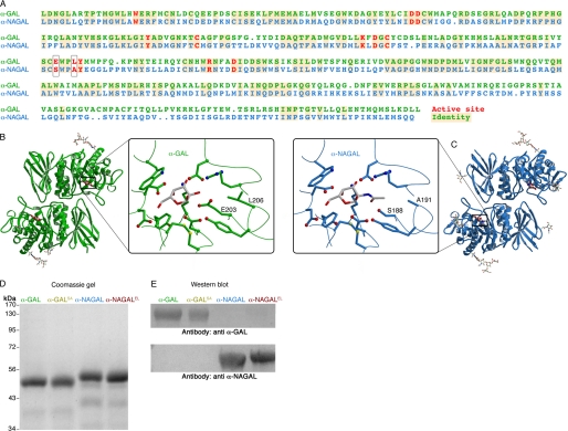
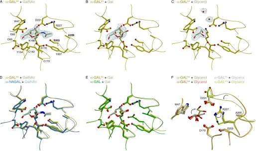
The human lysosomal enzymes alpha-galactosidase (alpha-GAL, EC 3.2.1.22) and alpha-N-acetylgalactosaminidase (alpha-NAGAL, EC 3.2.1.49) share 46% amino acid sequence identity and have similar folds. The active sites of the two enzymes share 11 of 13 amino acids, differing only where they interact with the 2-position of the substrates. Using a rational protein engineering approach, we interconverted the enzymatic specificity of alpha- GAL and alpha-NAGAL. The engineered alpha-GAL (which we call alpha-GAL(SA)) retains the antigenicity of alpha-GAL but has acquired the enzymatic specificity of alpha-NAGAL. Conversely, the engineered alpha-NAGAL (which we call alpha-NAGAL(EL)) retains the antigenicity of alpha-NAGAL but has acquired the enzymatic specificity of the alpha-GAL enzyme. Comparison of the crystal structures of the designed enzyme alpha-GAL(SA) to the wild-type enzymes shows that active sites of alpha-GAL(SA) and alpha-NAGAL superimpose well, indicating success of the rational design. The designed enzymes might be useful as non-immunogenic alternatives in enzyme replacement therapy for treatment of lysosomal storage disorders such as Fabry disease.
Figures
Similar articles
-
The 1.9 a structure of human alpha-N-acetylgalactosaminidase: The molecular basis of Schindler and Kanzaki diseases.J Mol Biol. 2009 Oct 23;393(2):435-47. doi: 10.1016/j.jmb.2009.08.021. Epub 2009 Aug 14. J Mol Biol. 2009. PMID: 19683538 Free PMC article.
-
The 1.9 A structure of alpha-N-acetylgalactosaminidase: molecular basis of glycosidase deficiency diseases.Structure. 2002 Mar;10(3):425-34. doi: 10.1016/s0969-2126(02)00726-8. Structure. 2002. PMID: 12005440
-
Structural basis of Fabry disease.Mol Genet Metab. 2002 Sep-Oct;77(1-2):3-11. doi: 10.1016/s1096-7192(02)00151-8. Mol Genet Metab. 2002. PMID: 12359124 Review.
-
The molecular defect leading to Fabry disease: structure of human alpha-galactosidase.J Mol Biol. 2004 Mar 19;337(2):319-35. doi: 10.1016/j.jmb.2004.01.035. J Mol Biol. 2004. PMID: 15003450
-
[Fabry-Anderson disease: current state of knowledge].Rev Invest Clin. 2011 May-Jun;63(3):314-21. Rev Invest Clin. 2011. PMID: 21888295 Review. Spanish.
Cited by
-
Human Alpha Galactosidases Transiently Produced in Nicotiana benthamiana Leaves: New Insights in Substrate Specificities with Relevance for Fabry Disease.Front Plant Sci. 2017 Jun 21;8:1026. doi: 10.3389/fpls.2017.01026. eCollection 2017. Front Plant Sci. 2017. PMID: 28680430 Free PMC article.
-
Venglustat, an orally administered glucosylceramide synthase inhibitor: Assessment over 3 years in adult males with classic Fabry disease in an open-label phase 2 study and its extension study.Mol Genet Metab. 2023 Feb;138(2):106963. doi: 10.1016/j.ymgme.2022.11.002. Epub 2022 Nov 9. Mol Genet Metab. 2023. PMID: 36481125 Free PMC article. Clinical Trial.
-
Carboxyl-terminal truncations alter the activity of the human α-galactosidase A.PLoS One. 2015 Feb 26;10(2):e0118341. doi: 10.1371/journal.pone.0118341. eCollection 2015. PLoS One. 2015. PMID: 25719393 Free PMC article.
-
Multiple exo-glycosidases in human serum as detected with the substrate DNP-α-GalNAc. I. A new assay for lysosomal α-N-acetylgalactosaminidase.BBA Clin. 2017 Oct 7;8:84-89. doi: 10.1016/j.bbacli.2017.10.001. eCollection 2017 Dec. BBA Clin. 2017. PMID: 29062717 Free PMC article.
-
Pharmacological chaperones for human α-N-acetylgalactosaminidase.Proc Natl Acad Sci U S A. 2012 Oct 23;109(43):17400-5. doi: 10.1073/pnas.1203924109. Epub 2012 Oct 8. Proc Natl Acad Sci U S A. 2012. PMID: 23045655 Free PMC article.
References
-
- Desnick R. J., Ioannou Y. A., Eng C. M. (2001) in The Metabolic and Molecular Bases of Inherited Disease (Scriver C. R., Beaudet A. L., Sly W. S., Valle D. eds) 8th Ed., pp. 3733–3774, McGraw-Hill, New York
-
- Desnick R. J., Schindler D. (2001) in The Metabolic and Molecular Bases of Inherited Disease (Scriver C. R., Beaudet A. L., Sly W. S., Valle D. eds) 8th Ed., pp. 3483–3505, McGraw-Hill, New York
-
- Wang A. M., Bishop D. F., Desnick R. J. (1990) J. Biol. Chem. 265, 21859–21866 - PubMed
Publication types
MeSH terms
Substances
Associated data
- Actions
- Actions
- Actions
- Actions
Grants and funding
LinkOut - more resources
Full Text Sources
Other Literature Sources
Miscellaneous
