. 2010 Jul 16;285(29):22448-60.
doi: 10.1074/jbc.M110.132274. Epub 2010 May 5.

Methylation of ribosomal protein L42 regulates ribosomal function and stress-adapted cell growth

Affiliations

Methylation of ribosomal protein L42 regulates ribosomal function and stress-adapted cell growth

Atsuko Shirai et al. J Biol Chem. .

Abstract

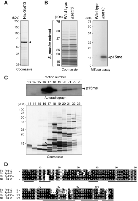
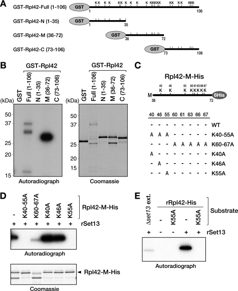
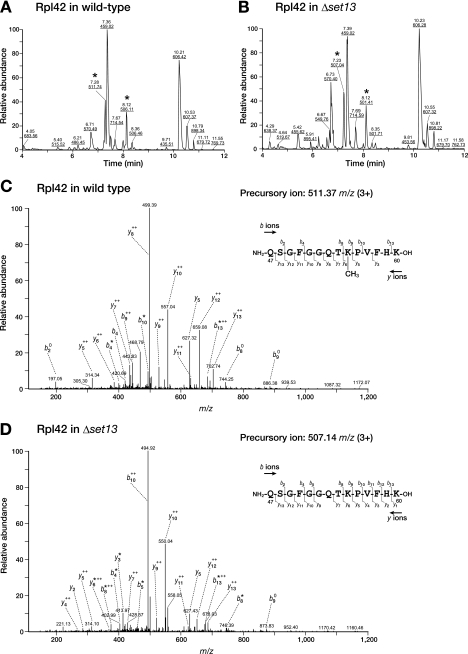
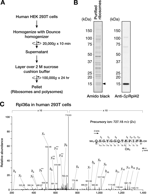
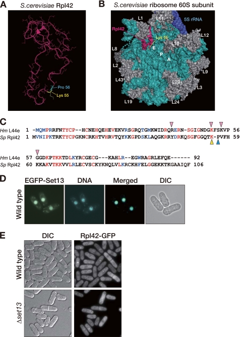
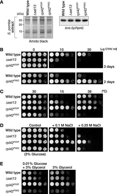
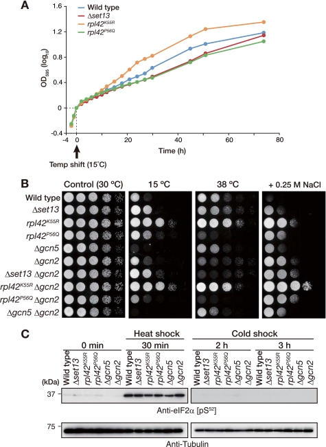
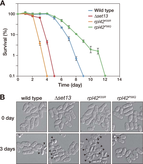
Lysine methylation is one of the most common protein modifications. Although lysine methylation of histones has been extensively studied and linked to gene regulation, that of non-histone proteins remains incompletely understood. Here, we show a novel regulatory role of ribosomal protein methylation. Using an in vitro methyltransferase assay, we found that Schizosaccharomyces pombe Set13, a SET domain protein encoded by SPAC688.14, specifically methylates lysine 55 of ribosomal protein L42 (Rpl42). Mass spectrometric analysis revealed that endogenous Rpl42 is monomethylated at lysine 55 in wild-type S. pombe cells and that the methylation is lost in Delta set13 mutant cells. Delta set13 and Rpl42 methylation-deficient mutant S. pombe cells showed higher cycloheximide sensitivity and defects in stress-responsive growth control compared with wild type. Genetic analyses suggested that the abnormal growth phenotype was distinct from the conserved stress-responsive pathway that modulates translation initiation. Furthermore, the Rpl42 methylation-deficient mutant cells showed a reduced ability to survive after entering stationary phase. These results suggest that Rpl42 methylation plays direct roles in ribosomal function and cell proliferation control independently of the general stress-response pathway.


Figures

FIGURE 1.
FIGURE 1.
S. pombe Set13 specifically methylates a protein in nuclear lysates. A, N-terminal His-tagged Set13 (His-Set13) was expressed in E. coli and purified by metal affinity chromatography. The eluted protein was resolved by 5–20% SDS-PAGE and visualized by Coomassie staining. His-Set13 is indicated by an arrowhead. B, in vitro MTase assay using S. pombe nuclear extracts. S. pombe nuclear extracts prepared from wild-type or Δset13 cells were resolved by 5–20% SDS-PAGE and visualized by Coomassie staining (left). These extracts were incubated with His-Set13 and [3H]AdoMet. The proteins were resolved by 15% SDS-PAGE, and labeled proteins were detected by autoradiography (right). The positions of size markers and labeled proteins are indicated, respectively, to the left and right of each image. C, fractionation of Set13 substrate(s) by reverse-phase chromatography. Nuclear extracts prepared from Δset13 cells were fractionated by reverse-phase chromatography. The proteins in each fraction were then concentrated and subjected to the in vitro MTase assay using His-Set13. After being resolved by 15% SDS-PAGE, the 3H-labeled proteins were detected by autoradiography (top), and the total proteins were detected by Coomassie staining (bottom). Protein(s) showing a similar migration profile to the 3H-labeled band is indicated by an arrowhead. D, amino acid sequences of S. pombe Rpl42 (Sp Rpl42), S. cerevisiae Rpl42 (Sc Rpl42), human Rpl36a (Hs Rpl36a), and Mus musculus Rpl44 (Mm Rpl44) aligned by the ClustalW 1.83 program. Identical amino acids are heavily shaded, and conserved amino acids are shown with light shading. The positions of peptide fragments identified in the LC-MS/MS analysis are indicated by black lines under the alignment.
FIGURE 2.
FIGURE 2.
Recombinant Rpl42 is methylated in vitro by His-Set13. A, schematic drawing of full-length and three fragments of Rpl42 as follows: the N terminus (GST-Rpl42-N), the middle part (GST-Rpl42-M), and the C terminus of the protein (GST-Rpl42-C). The position of each lysine residue is indicated by a K. The amino acid numbers are also shown. B, in vitro MTase assay using recombinant Rpl42. GST-Rpl42-Full, GST-Rpl42-N, GST-Rpl42-M, and GST-Rpl42-C were incubated with His-Set13 and [3H]AdoMet. The proteins were resolved by 15% SDS-PAGE and visualized by Coomassie staining (right). Proteins methylated by Set13 were detected by autoradiography (left). C, schematic representation of the middle part of Rpl42 (Rpl42-M-His) and lysine candidates for methylation. These eight lysine residues were replaced by nonmethylatable alanine either alone or in combination. A, alanine; WT, wild type. D and E, in vitro MTase assay using recombinant alanine-substituted mutants of Rpl42. Alanine-substituted mutants of Rpl42-M-His (D) and Rpl42-His (E) were incubated with His-Set13 and [3H]AdoMet. The proteins were resolved by 15% SDS-PAGE and visualized by Coomassie staining (D, bottom). Proteins methylated by Set13 were detected by autoradiography (D, top; E).
FIGURE 3.
FIGURE 3.
LC-MS/MS analysis of Rpl42 derived from wild-type and Δset13 S. pombe. Rpl42 was isolated from wild-type or Δset13 mutant cells by reverse-phase chromatography, digested with trypsin, and analyzed using a quadrupole ion trap mass spectrometer (Finnigan LTQ; Thermo Fisher Scientific). A and B, base peak, ion chromatogram for a 12-min separation of the digested Rpl42 peptides from the wild-type (A) and Δset13 strains (B). The elution time (upper) and representative m/z (lower, underlined) of the eluted peptides are indicated at the top of each peak. The peaks for the peptide fragment spanning residues 47–60 are indicated by an asterisk. C and D, MS/MS spectra of the peptide fragment spanning residues 47–60 from the wild-type (C) and Δset13 strains (D) are shown. The observed y and b ions and fragment map are shown.
FIGURE 4.
FIGURE 4.
Human Rpl36a is methylated at lysine 53 in vivo. A, procedure for preparing ribosomes and polysomes from HEK293T cells. B, detection of human Rpl36a using an anti-SpRpl42 antibody. Proteins were resolved by 8–16% SDS-PAGE and visualized by Coomassie staining (left). Rpl36a detected by the anti-SpRpl42 antibody is shown (right). The protein showing a similar migration as the Western blot signal is indicated by an arrowhead. C, LC-MS/MS analysis of human Rpl36a. Rpl36a excised from an SDS-polyacrylamide gel was digested with trypsin, and the digested peptides were subjected to LC-MS/MS analysis. The MS/MS spectrum of the peptide fragment spanning residues 45–57 is shown. The observed y and b ions and fragment map are shown.
FIGURE 5.
FIGURE 5.
Overview of Rpl42 on the large subunit of the ribosome. A, structure of S. cerevisiae Rpl42 (42). Blue, proline 56 of Rpl42; yellow, lysine 55 of Rpl42. B, surface view of the large subunit of the S. cerevisiae ribosome (Protein Data Bank file 1S1I) (42). Pink, Rpl42; aqua, 5.8 S/25 S ribosomal RNA; blue, 5 S ribosomal RNA; yellow, lysine 55 of Rpl42. The locations of the E-site, P-site, and A-site are indicated. The visible proteins are identified. C, amino acid sequences of S. pombe Rpl42 and H. marismortui L44e aligned by the ClustalW 2 program. Identical amino acids are in red, and conserved amino acids are in blue. Pink arrowheads indicate the main residues that interact with the 3′-CCA end of tRNA. Yellow arrowhead indicates the residue that is methylated in S. pombe Rpl42. Blue arrowhead indicates the residue whose substitution confers resistance to cycloheximide. D, EGFP-fused Set13 predominantly localized to the nucleus. EGFP-fused Set13 was expressed from the nmt1 promoter in wild-type cells. The cells were stained with Hoechst 33342 (DNA). A merged image of the EGFP-Set11 and DNA (Merged), and differential interference contrast (DIC) image are also shown. E, green fluorescent protein-fused Rpl42 mainly localized to the cytoplasm and the nucleolus. Rpl42-GFP was expressed from the nmt1 promoter in wild-type or Δset13 cells. The differential interference contrast image is also shown.
FIGURE 6.
FIGURE 6.
Rpl42 methylation-defective mutants show abnormal cell growth under environmental stresses. A, protein levels of Rpl42 in the Δset13, rpl42K55R, and rpl42P56Q mutant cells. Exponentially growing cultures of wild-type, Δset13, rpl42K55R, and rpl42P56Q cells in YEA were harvested. The total cell lysates prepared from these strains were resolved on a 12.5% SDS-polyacrylamide gel. Rpl42 was detected by Western blotting with an anti-SpRpl42 antibody (right). As a loading control, the Amido Black stained blot is shown (left). B, Δset13 and the rpl42K55R mutant cells showed a greater sensitivity to cycloheximide. 5-Fold dilutions of wild-type, Δset13, and rpl42K55R cells were plated onto YEA alone or YEA containing different doses of cycloheximide for 3 days (top). 5-Fold dilutions of wild-type and rpl42P56Q mutant cells were plated onto YEA alone or YEA containing different doses of cycloheximide for 2 days (bottom). C, set13-null and rpl42K55R mutant cells showed resistance against cold stress. 5-Fold dilutions of wild-type, Δset13, rpl42K55R, and rpl42P56Q cells were plated onto YEA at 30, 38, or 15 °C for 2, 3, or 19 days, respectively. D and E, rpl42K55R mutant cells showed resistance against environmental stresses. 5-Fold dilutions of each strain were plated onto YEA alone, YEA containing different doses of NaCl (D), YEA containing 3% glycerol instead of glucose, or YEA containing 0.01% glucose plus 3% glycerol (E), for 3 days.
FIGURE 7.
FIGURE 7.
Cold-adapted growth of Rpl42 methylation-defective mutants is independent of general stress-response pathways. A, effect of cold shock on cell growth. Wild-type (blue), Δset13 (red), rpl42K55R (orange), and rpl42P56Q (green) cells were grown until the mid-log phase in YEA medium at 30 °C. The cells were then shifted to 15 °C at time 0. Cell growth was monitored by measuring A595 (OD595). B, no genetic interaction between gcn2 and methylation-defective mutants was observed. 5-Fold dilutions of each strain were plated onto YEA containing 0.25 m NaCl for 4 days or YEA alone at 30, 38, or 15 °C for 2, 3, or 19 days, respectively. C, methylation-deficient mutation did not affect eIF2α phosphorylation in response to cold stress or heat shock. Exponentially growing cultures of wild-type, Δset13, rpl42K55R, rpl42P56Q, Δgcn5, and Δgcn2 cells growing in YEA were subjected to cold stress at 15 °C or heat shock at 48 °C for the times indicated. The total cell lysates prepared from the indicated strains were resolved on an 11% SDS-polyacrylamide gel. The level of eIF2a phosphorylation was analyzed by immunoblotting using an antibody that specifically recognizes phosphorylated eIF2α (top). The Western blot signal of tubulin was used as a loading control (bottom).
FIGURE 8.
FIGURE 8.
Rpl42 methylation and chronological life span. A, survival of wild-type, Δset13, rpl42K55A, and rpl42P56Q in stationary phase was evaluated by counting the colony-forming units. The y axis is shown in logarithmic scale. B, wild-type and mutant cells on the indicated days were observed under a light microscope. Arrowheads indicate abnormal or dead cells.

Similar articles

Cited by

References

    1. Clarke S. G., Tamanoi F. (eds) (2006) The Enzymes, 3rd Ed., Vol. 24, Academic Press, San Diego
    1. Qian C., Zhou M. M. (2006) Cell. Mol. Life Sci. 63, 2755–2763 - PMC - PubMed
    1. Lachner M., Jenuwein T. (2002) Curr. Opin. Cell Biol. 14, 286–298 - PubMed
    1. Martin C., Zhang Y. (2005) Nat. Rev. Mol. Cell Biol. 6, 838–849 - PubMed
    1. Klose R. J., Zhang Y. (2007) Nat. Rev. Mol. Cell Biol. 8, 307–318 - PubMed

Publication types

MeSH terms