. 2010 Jul 2;285(27):20850-9.
doi: 10.1074/jbc.M110.113910. Epub 2010 May 5.

Development of a two-part strategy to identify a therapeutic human bispecific antibody that inhibits IgE receptor signaling

Affiliations

Development of a two-part strategy to identify a therapeutic human bispecific antibody that inhibits IgE receptor signaling

Janet Jackman et al. J Biol Chem. .

Abstract

The development of bispecific antibodies as therapeutic agents for human diseases has great clinical potential, but broad application has been hindered by the difficulty of identifying bispecific antibody formats that exhibit favorable pharmacokinetic properties and ease of large-scale manufacturing. Previously, the development of an antibody technology utilizing heavy chain knobs-into-holes mutations and a single common light chain enabled the small-scale generation of human full-length bispecific antibodies. Here we have extended the technology by developing a two-part bispecific antibody discovery strategy that facilitates proof-of-concept studies and clinical candidate antibody generation. Our scheme consists of the efficient small-scale generation of bispecific antibodies lacking a common light chain and the hinge disulfides for proof-of-concept studies coupled with the identification of a common light chain bispecific antibody for large-scale production with high purity and yield. We have applied this technology to generate a bispecific antibody suitable for development as a human therapeutic. This antibody directly inhibits the activation of the high affinity IgE receptor FcepsilonRI on mast cells and basophils by cross-linking FcepsilonRI with the inhibitory receptor FcgammaRIIb, an approach that has strong therapeutic potential for asthma and other allergic diseases. Our approach for producing human bispecific full-length antibodies enables the clinical application of bispecific antibodies to a validated therapeutic pathway in asthma.


Figures

FIGURE 1.
FIGURE 1.
Expression and purification schemes for the generation of proof-of-concept and human therapeutic bispecific antibodies in E. coli. Proof-of-concept bispecific antibodies lacking a common light chain are produced at a small-scale as hingeless bispecific antibodies. Each arm of the bispecific antibody (knob and hole) is produced and purified separately. The two arms are then mixed together for annealing and subsequently purified and analyzed. Human therapeutic bispecific antibodies with a common light chain are produced in E. coli from a single plasmid containing knob and hole heavy chains and the common light chain. The intact bispecific antibody is subsequently purified and analyzed.
FIGURE 2.
FIGURE 2.
Generation and characterization of hingeless 22E7/5A6 bispecific antibody. A, shown is SDS-PAGE analysis of non-reduced 22E7/5A6 bispecific antibody shows high purity, with 22E7 hole and 5A6 knob co-migrating at an apparent molecular mass of ∼75 kDa as expected. Isoelectric focusing gel (IEF) analysis of the 22E7/5A6 bispecific antibody preparation indicates that it consists of a single heterodimeric antibody species with no contaminating 22E7 or 5A6 homodimers. Mass spectrometry analysis of non-reduced 22E7/5A6 bispecific antibody shows high purity and the expected masses of 22E7 hole and 5A6 knob. B, shown is ELISA analysis of 22E7/5A6 bispecific antibody binding to recombinant human FcϵRIα extracellular domain protein in the presence and absence of human IgE. C, shown is an ELISA analysis of 22E7/5A6 bispecific antibody binding to recombinant human FcγRIIb and FcγRIIa extracellular domains. D, shown is an ELISA analysis of 22E7/5A6 bispecific antibody, 5A6 knob, and 22E7 hole binding to both human FcϵRIα and human FcγRIIb extracellular domains. Recombinant human FcγRIIb is directly coated on the ELISA plate followed by incubation with 22E7/5A6, 22E7, or 5A6. Bispecific binding is detected by further incubation with recombinant human FcϵRIα extracellular domain followed by incubation with biotinylated human IgE binding and detection of the entire antibody/extracellular domain/IgE complex with HRP-conjugated streptavidin.
FIGURE 3.
FIGURE 3.
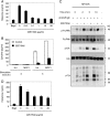
22E7/5A6 bispecific antibody inhibits mast cell and basophil activation. A, shown is the effect of 22E7/5A6 bispecific antibody on histamine release from RBL cells transfected with human FcϵRIα and human FcγRIIb and activated through human FcϵRI. B, shown is the effect of 22E7/5A6 bispecific antibody on interleukin-4 (IL4) and MCP-1 release from RBL cells transfected with human FcϵRIα and human FcγRIIb and activated through human FcϵRI. C, shown is a Western blot analysis of the effect of 22E7/5A6 bispecific antibody on phosphorylation of human FcγRIIb, ERK, and other protein tyrosines upon NP-ovalbumin (NP-Ova)-induced activation of NP-specific human IgE-sensitized RBL cells expressing human FcϵRIα and human FcγRIIb. D, shown is the effect of 22E7/5A6 bispecific antibody on histamine release from primary human basophils stimulated with polyclonal anti-IgE. Bkgd, background; MCP1, monocyte chemotactic protein 1.
FIGURE 4.
FIGURE 4.
22E7/14H6 bispecific antibody inhibits PCA. Dermal mast cells of human FcϵRIα transgenic mice were passively sensitized with NP-specific human IgE 24 h before activation. 22E7/14H6 bispecific antibody was administered after cutaneous mast cell sensitization, 2 h before mast cell activation. Mast cells were activated by intravenous injection of 200 μg NP-ovalbumin with 1% Evans blue. A, shown are pictures of skin from human FcϵRIα transgenic mice treated with control or 22E7/14H6 bispecific antibody and subjected to PCA. B, shown is quantitation of Evans blue dye extracted from the flank skin of human FcϵRIα transgenic mice treated with 22E7/14H6 antibody and subjected to PCA. *, p < 0.05, compared with control antibody treatment.
FIGURE 5.
FIGURE 5.
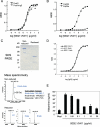
Generation and characterization of 9202.1/5411 bispecific antibody. A, shown is an ELISA analysis of 9202.1/5411 bispecific antibody binding to recombinant human FcϵRIα extracellular domain protein in the presence and absence of human IgE. B, shown is an ELISA analysis of 9202.1/5411 bispecific antibody binding to recombinant human FcγRIIb and FcγRIIa extracellular domains. C, shown is SDS-PAGE analysis of 9202.1/5411 bispecific antibody under non-reducing and reducing conditions indicates high purity and the expected masses of heavy and light chains (LC). Mass spectrometry analysis of 9202.1/5411 bispecific antibody (non-reduced and reduced) indicates expected masses of the intact bispecific antibody, common light chain, 9202.1 hole, and 5411 knob. D, shown is an ELISA analysis of 9202.1/5411 bispecific antibody, 5411 knob, and 9202.1 hole binding to both human FcϵRIα and human FcγRIIb extracellular domains. Recombinant human FcγRIIb is directly coated on the ELISA plate followed by incubation with 9202.1/5411, 9202.1, or 5411. Bispecific binding is detected by further incubation with recombinant human FcϵRIα extracellular domain followed by incubation with biotinylated human IgE binding and detection of the entire antibody/extracellular domain/IgE complex with HRP-conjugated streptavidin. E, shown is the effect of 9202.1/5411 bispecific antibody on histamine release from RBL cells transfected with human FcϵRIα and human FcγRIIb and activated through human FcϵRI. Bkgd, background.

Similar articles

Cited by

References

    1. Bargou R., Leo E., Zugmaier G., Klinger M., Goebeler M., Knop S., Noppeney R., Viardot A., Hess G., Schuler M., Einsele H., Brandl C., Wolf A., Kirchinger P., Klappers P., Schmidt M., Riethmüller G., Reinhardt C., Baeuerle P. A., Kufer P. (2008) Science 321, 974–977 - PubMed
    1. van Spriel A. B., van Ojik H. H., van De Winkel J. G. (2000) Immunol. Today 21, 391–397 - PubMed
    1. Asano R., Watanabe Y., Kawaguchi H., Fukazawa H., Nakanishi T., Umetsu M., Hayashi H., Katayose Y., Unno M., Kudo T., Kumagai I. (2007) J. Biol. Chem. 282, 27659–27665 - PubMed
    1. Carter P. (2001) J. Immunol. Methods 248, 7–15 - PubMed
    1. Kufer P., Lutterbüse R., Baeuerle P. A. (2004) Trends Biotechnol. 22, 238–244 - PubMed

Publication types

MeSH terms