. 2010 Jul 9;285(28):21849-57.
doi: 10.1074/jbc.M110.141010. Epub 2010 May 5.

Cdk1 activity is required for mitotic activation of aurora A during G2/M transition of human cells

Affiliations

Cdk1 activity is required for mitotic activation of aurora A during G2/M transition of human cells

Robert D Van Horn et al. J Biol Chem. .

Abstract

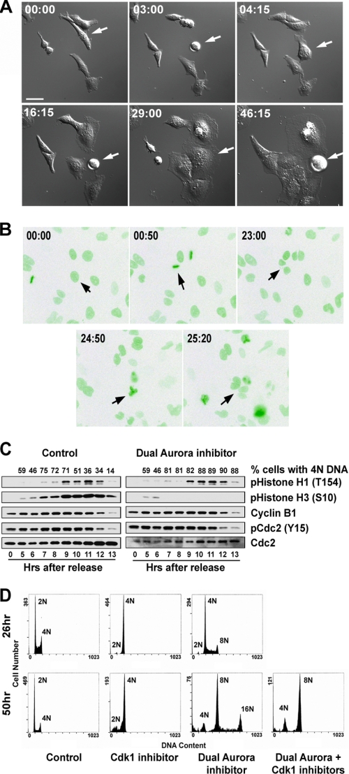
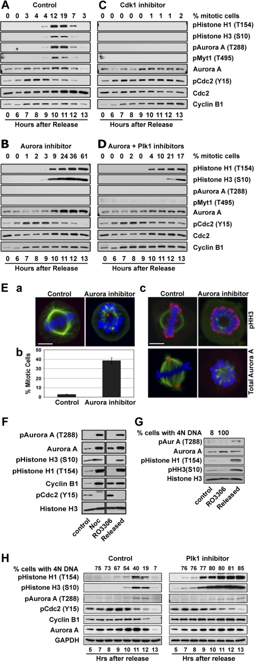
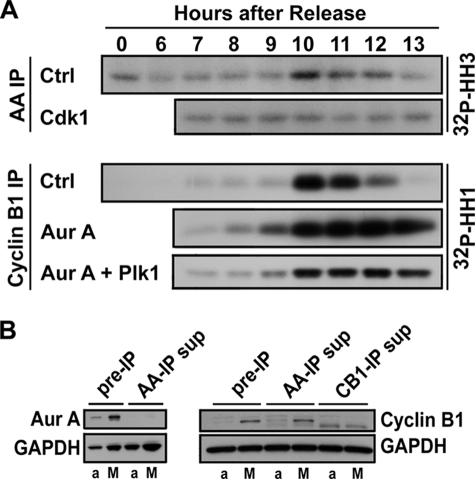
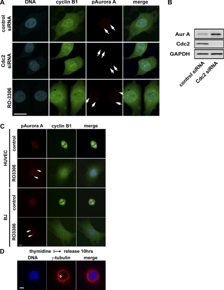
In mammalian cells entry into and progression through mitosis are regulated by multiple mitotic kinases. How mitotic kinases interact with each other and coordinately regulate mitosis remains to be fully understood. Here we employed a chemical biology approach using selective small molecule kinase inhibitors to dissect the relationship between Cdk1 and Aurora A kinases during G(2)/M transition. We find that activation of Aurora A first occurs at centrosomes at late G(2) and is required for centrosome separation independently of Cdk1 activity. Upon entry into mitosis, Aurora A then becomes fully activated downstream of Cdk1 activation. Inactivation of Aurora A or Plk1 individually during a synchronized cell cycle shows no significant effect on Cdk1 activation and entry into mitosis. However, simultaneous inactivation of both Aurora A and Plk1 markedly delays Cdk1 activation and entry into mitosis, suggesting that Aurora A and Plk1 have redundant functions in the feedback activation of Cdk1. Together, our data suggest that Cdk1, Aurora A, and Plk1 mitotic kinases participate in a feedback activation loop and that activation of Cdk1 initiates the feedback loop activity, leading to rapid and timely entry into mitosis in human cells. In addition, live cell imaging reveals that the nuclear cycle of cells becomes uncoupled from cytokinesis upon inactivation of both Aurora A and Aurora B kinases and continues to oscillate in a Cdk1-dependent manner in the absence of cytokinesis, resulting in multinucleated, polyploidy cells.


Figures

FIGURE 1.
FIGURE 1.
Effects of inactivation of Aurora kinases on cell cycle progression. Repeated nuclear division without cytokinesis in the presence of a dual Aurora kinase inhibitor revealed by live cell imaging is shown. A, shown are snapshots of differential interference contrast images of HeLa cells undergoing repeated rounds of mitosis without the ensuing cytokinesis in the presence of a dual Aurora kinase inhibitor. The arrow points to a cell that underwent three rounds of mitosis with increasing cell size. B, shown are snapshots of fluorescent green fluorescent protein images of U2OS cells expressing green fluorescent protein-H2B in the presence of a dual Aurora kinase inhibitor. The same cell that underwent more than two rounds of mitosis is indicated by arrows in each image. The time after the addition of the dual Aurora kinase inhibitor when the image was acquired is indicated in each image. Increased size of the cells as a function of time after the dual Aurora kinase inhibitor addition (panel A) was noted. Bar = 40 μm. C, shown is a Western blot analysis of a synchronized mitosis in the presence or absence of Aurora kinase activities. Cells were synchronized by double thymidine block/release, and the dual Aurora kinase inhibitor, ZM44739, was added 5 h after thymidine release. The numbers on the top represent the % of cells with 4N DNA. D, nuclear division in the absence of Aurora kinase activities is Cdk1-dependent. DNA contents of synchronized HeLa S3 cells at the times indicated after treatment with a selective Cdk1 kinase inhibitor or a dual Aurora kinase inhibitor or a sequential treatment with a dual Aurora kinase inhibitor first for 24 h followed by a selective Cdk1 kinase inhibitor.
FIGURE 2.
FIGURE 2.
Characterization of a Cdk1-dependent phospho-specific histone H1 (Thr(P)-154) antibody. A, shown is Western analysis of phospho-histone H1 antibody (Thr(P)-154) showing specific cross-reactivity with mitotically phosphorylated histone H1. The loading of the gel was as follows: lane 1, molecular weight marker; lane 2, 1 μg of control unphosphorylated histone H1; lane 3, 1 μg of histone H1 isolated from colcemid-treated HeLa cells; lane 4, 28 μg of protein lysates from CaLu-6 cells; lane 5, 28 μg of protein lysates from CaLu-6 cells treated with nocodazole. B, shown is specific staining of mitotic cells by the phospho-specific histone H1 antibody (Thr(P)-154). Blue = DNA; green = phospho-histone H1; red = α-tubulin. Bar = 60 μm. C, shown is a Western blot analysis of histone H1 in vitro phosphorylated by Cdk1 with the phospho-specific histone H1 antibody (Thr(P)-154). Lanes 1 and 2, Cdk1 kinase alone, from UBI and New England BioLabs, respectively; lane 2, unphosphorylated histone H1 protein control; lanes 4 and 5, histone H1 protein phosphorylated in vitro by Cdk1 kinase from UBI and New England BioLabs, respectively. The upper panel is the Western blot, and the lower panel is Coomassie staining of the gel showing equal loading of histone H1 for Western blot analysis.
FIGURE 3.
FIGURE 3.
Aurora kinases are not required for entry into mitosis and activation of Cdk1. A, HeLa cells were synchronized by double thymidine block/release. Synchronized cells were treated with DMSO as control (A) or a selective Aurora A kinase inhibitor (B) or a selective Cdk1 kinase inhibitor (C) or a combination of Aurora A and Plk1 kinase inhibitors (D) 5 h after release from thymidine block. Mitotic index was analyzed by flow cytometry and mitotic progression was also followed biochemically by Western blotting for phospho-histone H1 and H3, autophosphorylation of Aurora A (Thr(P)-288), Myt1 phosphorylation at Thr-495, Cdc2 tyrosine 15 phosphorylation (Y15) and cyclin B1 and Aurora A protein levels. Total Cdc2 protein was used as loading control. E, shown is mitotic arrest with monopolar spindles of cells treated with a selective Aurora A kinase inhibitor. a, shown is a representative control and a treated HeLa cell showing normal bipolar mitotic spindle in the control and monopolar spindle in the treated cell. Blue = DNA; red = pericentrin staining of centrosome; green = α-tubulin staining. b, shown is the mitotic index of the control and cells treated with a selective Aurora kinase inhibitor, which was used at a concentration shown to selectively inhibit Aurora A kinase activity. Cells were fixed and stained as shown in panel A. At least 500 cells were counted, and mitotic index is expressed as percentage of cells at mitosis. c, shown is a mitotic control and a treated cell showing phospho-histone H3 and Aurora A staining. Blue = DNA; green = α-tubulin; red = phospho-histone H3 (upper) and Aurora A (lower). Note that the Aurora A kinase inhibitor prevented centrosome localization of the Aurora A protein. Bar = 7 μm. F, Cdk1 inhibitor, RO3306 (9 μm), block/release of HeLa cells is shown. Cells were treated with RO3306 for 20 h and then were released from the block by washing 3× with fresh medium without RO3306. Total cell lysates were analyzed by Western blot analysis for mitotic markers and activation of Aurora A kinase. Mitotic extract of cells treated with nocodazole for 20 h was used as a positive control. G, NIH-3T3 cells were similarly treated with RO3306 as described for panel F for Cdk1-dependent activation of Aurora A kinase during G2/M transition. H, shown is Western blot analysis of a synchronized cell cycle progression in the presence or absence of a selective Plk1 kinase inhibitor, BI2536. HeLa cells were synchronized by double thymidine block, and the Plk1 inhibitor was added 5 h after release from thymidine block. GAPDH, glyceraldehyde-3-phosphate dehydrogenase.
FIGURE 4.
FIGURE 4.
In vitro kinase assays. HeLa cells were synchronized by double thymidine block/release and were treated with the kinase inhibitors as described for Fig. 3. A, Cdk1 and Aurora A kinases were immunoprecipitated (IP) using cyclin B1 and Aurora A antibodies, and their kinase activities were assed in vitro with histone H1 (HH1) and histone H3 (HH3) as substrates, respectively. Ctrl, control. B, effective depletion of Aurora A (Aur A) and cyclin B1 by immunoprecipitation is shown. An asynchronous growing cell sample (a) and a mitotic cell sample (M) were incubated with Aurora A (AA-IP)- and cyclin B1 (CB1-IP)-specific antibodies, respectively, under the same conditions for kinase assays by IP as shown in panel A. After removal of the IP complex, the supernatants (sup) were analyzed by Western blotting for completeness of target protein depletion by immunoprecipitation. Please note that immunoprecipitation completely depleted Aurora A and cyclin B1 from both asynchronous and mitotic cells samples. GAPDH, glyceraldehyde-3-phosphate dehydrogenase.
FIGURE 5.
FIGURE 5.
Cdk1-independent activation of Aurora A at centrosomes and centrosome separation at G2. A, representative images of HeLa cells treated with the small molecule Cdk1 inhibitor, RO3306 (9 μm), or Cdk1-specific siRNA and stained for cyclin B1, phospho-specific Aurora A (Thr-288), and DNA are shown. Bar = 20 μm. B, Western blots show effective knockdown of Cdc2 proteins by the Cdc2-specific siRNA oligo. Accumulation of Aurora A (Aur A) kinase in the absence of Cdk1 activity is consistent with G2 cell cycle arrest. GAPDH, glyceraldehyde-3-phosphate dehydrogenase. C, shown are representative images of human umbilical vein endothelial (HUVEC) and BJ cells, non-transformed human cells treated with the Cdk1 kinase inhibitor RO3306 (9 μm) and stained for cyclin B1, and phospho-specific Aurora A (pAurora A; Thr-288) and DNA. Bar = 10 μm. D, shown are representative images of HeLa cells treated with Cdk1 and Aurora kinase inhibitors as indicated and stained for DNA and γ-tubulin. Bar = 10 μm. Cells with inactivated Cdk1 were cleanly arrested at G2. Several hundred cells were examined under the microscope, and all had cytoplasmic cyclin B1 accumulation and activated Aurora A kinase localized to centrosomes which are well separated.

Similar articles

Cited by

References

    1. Nigg E. A. (2001) Nat. Rev. Mol. Cell Biol. 2, 21–32 - PubMed
    1. Nurse P. (1994) Cell 79, 547–550 - PubMed
    1. Osmani S. A., Ye X. S. (1996) Biochem. J. 317, 633–641 - PMC - PubMed
    1. Ye X. S., Xu G., Pu R. T., Fincher R. R., McGuire S. L., Osmani A. H., Osmani S. A. (1995) EMBO J. 14, 986–994 - PMC - PubMed
    1. Qian Y. W., Erikson E., Taieb F. E., Maller J. L. (2001) Mol. Biol. Cell 12, 1791–1799 - PMC - PubMed

LinkOut - more resources