

The Trypanosoma brucei life cycle switch TbPTP1 is structurally conserved and dephosphorylates the nucleolar protein NOPP44/46
- PMID: 20444707
- PMCID: PMC2903352
- DOI: 10.1074/jbc.M110.108860
The Trypanosoma brucei life cycle switch TbPTP1 is structurally conserved and dephosphorylates the nucleolar protein NOPP44/46
Abstract
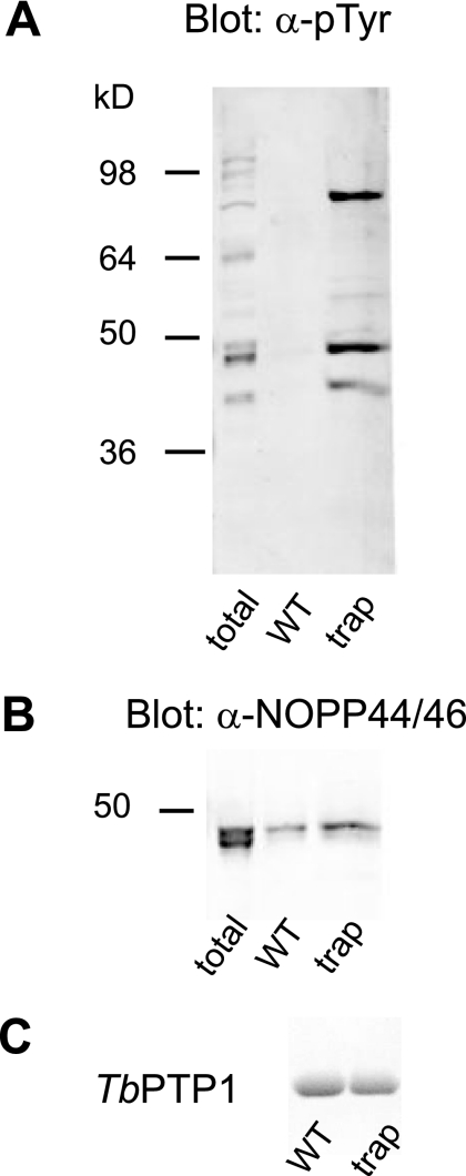
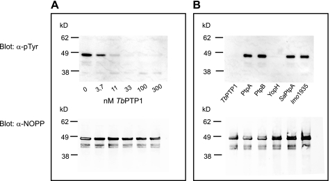
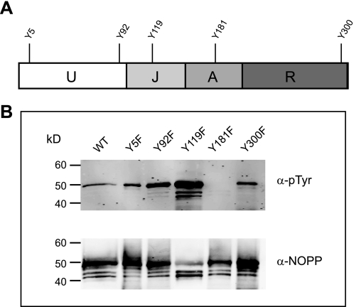
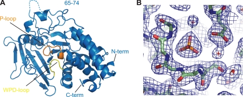
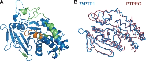
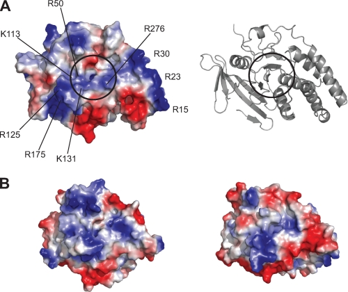
Trypanosoma brucei adapts to changing environments as it cycles through arrested and proliferating stages in the human and tsetse fly hosts. Changes in protein tyrosine phosphorylation of several proteins, including NOPP44/46, accompany T. brucei development. Moreover, inactivation of T. brucei protein-tyrosine phosphatase 1 (TbPTP1) triggers differentiation of bloodstream stumpy forms into tsetse procyclic forms through unknown downstream effects. Here, we link these events by showing that NOPP44/46 is a major substrate of TbPTP1. TbPTP1 substrate-trapping mutants selectively enrich NOPP44/46 from procyclic stage cell lysates, and TbPTP1 efficiently and selectively dephosphorylates NOPP44/46 in vitro. To provide insights into the mechanism of NOPP44/46 recognition, we determined the crystal structure of TbPTP1. The TbPTP1 structure, the first of a kinetoplastid protein-tyrosine phosphatase (PTP), emphasizes the conservation of the PTP fold, extending to one of the most diverged eukaryotes. The structure reveals surfaces that may mediate substrate specificity and affords a template for the design of selective inhibitors to interfere with T. brucei transmission.
Figures
Similar articles
-
Protein tyrosine phosphatase TbPTP1: A molecular switch controlling life cycle differentiation in trypanosomes.J Cell Biol. 2006 Oct 23;175(2):293-303. doi: 10.1083/jcb.200605090. Epub 2006 Oct 16. J Cell Biol. 2006. PMID: 17043136 Free PMC article.
-
Positional Dynamics and Glycosomal Recruitment of Developmental Regulators during Trypanosome Differentiation.mBio. 2019 Jul 9;10(4):e00875-19. doi: 10.1128/mBio.00875-19. mBio. 2019. PMID: 31289175 Free PMC article.
-
A novel phosphatase cascade regulates differentiation in Trypanosoma brucei via a glycosomal signaling pathway.Genes Dev. 2010 Jun 15;24(12):1306-16. doi: 10.1101/gad.570310. Genes Dev. 2010. PMID: 20551176 Free PMC article.
-
Molecular cloning of Trypanosoma brucei CK2 catalytic subunits: the alpha isoform is nucleolar and phosphorylates the nucleolar protein Nopp44/46.Mol Biochem Parasitol. 2002 Jan;119(1):97-106. doi: 10.1016/s0166-6851(01)00407-8. Mol Biochem Parasitol. 2002. PMID: 11755190
-
A paradigm shift: The mitoproteomes of procyclic and bloodstream Trypanosoma brucei are comparably complex.PLoS Pathog. 2017 Dec 21;13(12):e1006679. doi: 10.1371/journal.ppat.1006679. eCollection 2017 Dec. PLoS Pathog. 2017. PMID: 29267392 Free PMC article. Review. No abstract available.
Cited by
-
The Potential of Secondary Metabolites from Plants as Drugs or Leads against Protozoan Neglected Diseases-Part III: In-Silico Molecular Docking Investigations.Molecules. 2016 Oct 19;21(10):1389. doi: 10.3390/molecules21101389. Molecules. 2016. PMID: 27775577 Free PMC article. Review.
-
Allostery in Protein Tyrosine Phosphatases is Enabled by Divergent Dynamics.J Chem Inf Model. 2024 Feb 26;64(4):1331-1346. doi: 10.1021/acs.jcim.3c01615. Epub 2024 Feb 12. J Chem Inf Model. 2024. PMID: 38346324 Free PMC article.
-
Structure of the Trypanosoma cruzi protein tyrosine phosphatase TcPTP1, a potential therapeutic target for Chagas' disease.Mol Biochem Parasitol. 2013 Jan;187(1):1-8. doi: 10.1016/j.molbiopara.2012.10.006. Epub 2012 Nov 5. Mol Biochem Parasitol. 2013. PMID: 23137716 Free PMC article.
-
Trypanosomatid protein phosphatases.Mol Biochem Parasitol. 2010 Oct;173(2):53-63. doi: 10.1016/j.molbiopara.2010.05.017. Epub 2010 Jun 1. Mol Biochem Parasitol. 2010. PMID: 20594956 Free PMC article. Review.
-
Inhibition of Trypanosoma evansi Protein-Tyrosine Phosphatase by Myristic Acid Analogues Isolated from Khaya senegalensis and Tamarindus indica.J Exp Pharmacol. 2019 Dec 18;11:135-148. doi: 10.2147/JEP.S226632. eCollection 2019. J Exp Pharmacol. 2019. PMID: 31908547 Free PMC article.
References
-
- Brun R., Blum J., Chappuis F., Burri C. (2010) Lancet 375, 148–159 - PubMed
-
- Parsons M., Valentine M., Deans J., Schieven G. L., Ledbetter J. A. (1991) Mol. Biochem. Parasitol. 45, 241–248 - PubMed
-
- García-Salcedo J. A., Nolan D. P., Gijón P., Gómez-Rodriguez J., Pays E. (2002) Mol. Microbiol. 45, 307–319 - PubMed
Publication types
MeSH terms
Substances
Associated data
- Actions
Grants and funding
LinkOut - more resources
Full Text Sources
