

Review
doi: 10.1152/ajprenal.00174.2010.
Epub 2010 May 5.
Hypoxic regulation of erythropoiesis and iron metabolism
Affiliations
- PMID: 20444740
- PMCID: PMC2904169
- DOI: 10.1152/ajprenal.00174.2010
Item in Clipboard
Review
Hypoxic regulation of erythropoiesis and iron metabolism
Am J Physiol Renal Physiol.
2010 Jul.
Abstract
The kidney is a highly sensitive oxygen sensor and plays a central role in mediating the hypoxic induction of red blood cell production. Efforts to understand the molecular basis of oxygen-regulated erythropoiesis have led to the identification of erythropoietin (EPO), which is essential for normal erythropoiesis and to the purification of hypoxia-inducible factor (HIF), the transcription factor that regulates EPO synthesis and mediates cellular adaptation to hypoxia. Recent insights into the molecular mechanisms that control and integrate cellular and systemic erythropoiesis-promoting hypoxia responses and their potential as a therapeutic target for the treatment of renal anemia are discussed in this review.
Figures
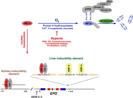
Hypoxia-inducible factor (HIF)-2 regulates erythropoietin (EPO). Shown is an overview of EPO gene regulation by the von Hippel-Lindau (VHL)/HIF/prolyl-4-hydroxylase domain (PHD) oxygen-sensing pathway. Proteasomal degradation of HIF-2α by the VHL tumor suppressor (pVHL)-E3-ubiquitin ligase complex (shown are key components of this complex) requires hydroxylation by oxygen- and iron-dependent PHDs. Binding to hydroxylated HIF-α occurs at the β-domain of pVHL, which spans amino acid residues 64–154. The C-terminal α-domain links the substrate recognition component pVHL to the E3 ubiquitin ligase via elongin C. In the absence of molecular oxygen, HIF-2α is not degraded and translocates to the nucleus where it forms a heterodimer with HIF-β, also known as the aryl hydrocarbon receptor nuclear translocator (ARNT). HIF-2α/β heterodimers bind to the HIF consensus binding site 5′-RCGTG-3′ and increase EPO transcription in the presence of transcriptional coactivators, such as CREB-binding protein (CBP) and p300. Hypoxic induction of EPO in the liver is mediated by the liver-inducibility element located in the 3′-end of the EPO gene and in renal interstitial fibroblast-like cells by the 5′-kidney-inducibility element, which is located 6–14 kb upstream of its transcription start site. Nitric oxide, reactive oxygen species, Krebs cycle metabolites succinate and fumarate, cobalt chloride (CoCl2), and iron chelators such as desferrioxamine inhibit HIF PHDs in the presence of oxygen, resulting in increased EPO transcription. EPO mRNA is encoded by 5 exons depicted by boxes. Coding sequences are shown in red. Nontranslated regions are shown in blue, and numbers indicate distance from the transcription start site in kb (not drawn to scale). Also shown are binding sites for hepatocyte nuclear factor (HNF)-4 in the 3′-liver-inducibility region. Fe2+, ferrous iron; NO, nitric oxide; ROS, reactive oxygen species; ub, ubiquitin.
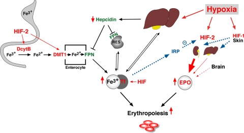
Hypoxia coordinates EPO synthesis with iron metabolism. Shown is a simplified overview of hypoxic and HIF-mediated effects on iron metabolism. HIF-2 induces renal and hepatic EPO synthesis in response to hypoxia, which results in increased serum EPO levels (circle), stimulating erythropoiesis. Renal and liver EPO responses are modulated by dermal HIF-1 (see the text). Also, included in this schematic is the recently described contribution of glial cell-derived EPO to the serum EPO pool. An adjustment of iron metabolism is needed to satisfy increased iron demand in the bone marrow. In the duodenum, duodenal cytochrome b (DcytB) reduces ferric iron (Fe3+) to its ferrous form (Fe2+), which is then transported into the cytosol of enterocytes (square) by divalent metal transporter-1 (DMT1). DcytB and DMT1 are both hypoxia inducible and HIF-2 regulated. Absorbed iron is released into the circulation by ferroportin (FPN) and is then transported in complex with transferrin to liver, reticuloendothelial cells, bone marrow, and other organs. Transferrin (Tf) is HIF regulated, and hypoxia increases its serum levels. Hypoxia, low serum iron levels, and increased “erythropoietic drive” inhibit hepcidin synthesis in the liver, resulting in diminished FPN cell surface expression in different tissues. As a result, more iron is released from enterocytes, hepatocytes, and reticuloendothelial cells (RES). When intracellular iron levels are low, iron regulatory protein (IRP) inhibits HIF-2α translation and diminishes hypoxia-induced erythropoiesis.
References
-
- An WG, Kanekal M, Simon MC, Maltepe E, Blagosklonny MV, Neckers LM. Stabilization of wild-type p53 by hypoxia-inducible factor 1alpha. Nature 392: 405–408, 1998 - PubMed
-
- Ang SO, Chen H, Hirota K, Gordeuk VR, Jelinek J, Guan Y, Liu E, Sergueeva AI, Miasnikova GY, Mole D, Maxwell PH, Stockton DW, Semenza GL, Prchal JT. Disruption of oxygen homeostasis underlies congenital Chuvash polycythemia. Nat Genet 32: 614–621, 2002 - PubMed
Publication types
MeSH terms
Substances
Grants and funding
LinkOut - more resources
Full Text Sources
Other Literature Sources
Medical
Research Materials