

Kidney-specific inactivation of Ofd1 leads to renal cystic disease associated with upregulation of the mTOR pathway
- PMID: 20444807
- PMCID: PMC2893811
- DOI: 10.1093/hmg/ddq180
Kidney-specific inactivation of Ofd1 leads to renal cystic disease associated with upregulation of the mTOR pathway
Abstract
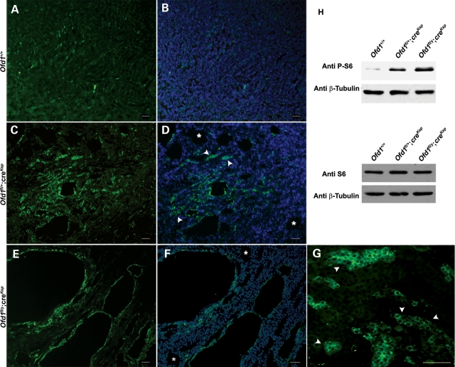
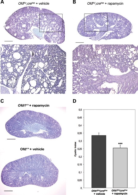
The oral-facial-digital type I syndrome (OFDI; MIM 311200) is a rare syndromic form of inherited renal cystic disease. It is transmitted as an X-linked dominant, male lethal disorder and is caused by mutations in the OFD1 gene. Previous studies demonstrated that OFDI belongs to the growing number of disorders ascribed to dysfunction of primary cilia. We generated a conditional inactivation of the mouse Ofd1 gene using the Ksp-Cre transgenic line, which resulted in a viable model characterized by renal cystic disease and progressive impairment of renal function. The study of this model allowed us to demonstrate that primary cilia initially form and then disappear after the development of cysts, suggesting that the absence of primary cilia is a consequence rather than the primary cause of renal cystic disease. Immunofluorescence and western blotting analysis revealed upregulation of the mTOR pathway in both dilated and non-dilated renal structures. Treatment with rapamycin, a specific inhibitor of the mTOR pathway, resulted in a significant reduction in the number and size of renal cysts and a decrease in the cystic index compared with untreated mutant animals, suggesting that dysregulation of this pathway in our model is mTOR-dependent. The animal model we have generated could thus represent a valuable tool to understand the molecular link between mTOR and cyst development, and eventually to the identification of novel drug targets for renal cystic disease.
Figures

Similar articles
-
Loss of primary cilia upregulates renal hypertrophic signaling and promotes cystogenesis.J Am Soc Nephrol. 2011 May;22(5):839-48. doi: 10.1681/ASN.2010050526. Epub 2011 Apr 14. J Am Soc Nephrol. 2011. PMID: 21493775 Free PMC article.
-
Mammalian target of rapamycin regulates vascular endothelial growth factor-dependent liver cyst growth in polycystin-2-defective mice.Hepatology. 2010 May;51(5):1778-88. doi: 10.1002/hep.23511. Hepatology. 2010. PMID: 20131403 Free PMC article.
-
Failure to ubiquitinate c-Met leads to hyperactivation of mTOR signaling in a mouse model of autosomal dominant polycystic kidney disease.J Clin Invest. 2010 Oct;120(10):3617-28. doi: 10.1172/JCI41531. Epub 2010 Sep 13. J Clin Invest. 2010. PMID: 20852388 Free PMC article.
-
The molecular basis of oral-facial-digital syndrome, type 1.Am J Med Genet C Semin Med Genet. 2009 Nov 15;151C(4):318-25. doi: 10.1002/ajmg.c.30224. Am J Med Genet C Semin Med Genet. 2009. PMID: 19876934 Review.
-
Cilia and centrosomes: a unifying pathogenic concept for cystic kidney disease?Nat Rev Genet. 2005 Dec;6(12):928-40. doi: 10.1038/nrg1727. Nat Rev Genet. 2005. PMID: 16341073 Review.
Cited by
-
Functional Genomics of the Retina to Elucidate its Construction and Deconstruction.Int J Mol Sci. 2019 Oct 4;20(19):4922. doi: 10.3390/ijms20194922. Int J Mol Sci. 2019. PMID: 31590277 Free PMC article. Review.
-
IK is essentially involved in ciliogenesis as an upstream regulator of oral-facial-digital syndrome ciliopathy gene, ofd1.Cell Biosci. 2023 Oct 28;13(1):195. doi: 10.1186/s13578-023-01146-9. Cell Biosci. 2023. PMID: 37898820 Free PMC article.
-
Signaling through the Primary Cilium.Front Cell Dev Biol. 2018 Feb 8;6:8. doi: 10.3389/fcell.2018.00008. eCollection 2018. Front Cell Dev Biol. 2018. PMID: 29473038 Free PMC article. Review.
-
Meckel-Gruber syndrome and the role of primary cilia in kidney, skeleton, and central nervous system development.Organogenesis. 2014 Jan 1;10(1):96-107. doi: 10.4161/org.27375. Epub 2013 Dec 9. Organogenesis. 2014. PMID: 24322779 Free PMC article. Review.
-
The HOPS complex subunit VPS39 controls ciliogenesis through autophagy.Hum Mol Genet. 2020 Apr 15;29(6):1018-1029. doi: 10.1093/hmg/ddaa029. Hum Mol Genet. 2020. PMID: 32077937 Free PMC article.
References
-
- Franco B. The molecular basis of oral-facial-digital type I (OFDI) syndrome. In: Epstein J.C., Erickson R.P., Wynshaw-Boris A., editors. Inborn Errors of Development. 2nd edn. New York: Oxford University Press; 2008. pp. 1379–1386. Chapter 156, Vol. I.
-
- Coll E., Torra R., Pascual J., Botey A., Ara J., Perez L., Ballesta F., Darnell A. Sporadic orofaciodigital syndrome type 1 presenting as end-stage renal disease. Nephrol. Dial. Transplant. 1997;12:1040–1042. doi:10.1093/ndt/12.5.1040. - DOI - PubMed
-
- Feather S.A., Winyard P.J., Dodd S., Woolf A.S. Oral-facial-digital syndrome type 1 is another dominant polycystic kidney disease: clinical, radiological and histopathological features of a new kindred. Nephrol. Dial. Transplant. 1997;12:1354–1361. doi:10.1093/ndt/12.7.1354. - DOI - PubMed
-
- Prattichizzo C., Macca M., Novelli V., Giorgio G., Barra A., Franco B. Mutational spectrum of the oral-facial-digital type I syndrome: a study on a large collection of patients. Hum. Mutat. 2008;29:1237–1246. doi:10.1002/humu.20792. - DOI - PubMed
-
- Doege T., Thuline H., Priest J., Norby D., Bryant J. Studies of a family with the oral-facial-digital syndrome. N. Engl. J. Med. 1964;271:1073–1080. - PubMed
Publication types
MeSH terms
Substances
Grants and funding
LinkOut - more resources
Full Text Sources
Medical
Molecular Biology Databases
Miscellaneous
