. 2010 Aug;84(15):7459-72.
doi: 10.1128/JVI.01875-09. Epub 2010 May 5.

Processing of lagging-strand intermediates in vitro by herpes simplex virus type 1 DNA polymerase

Affiliations

Processing of lagging-strand intermediates in vitro by herpes simplex virus type 1 DNA polymerase

Yali Zhu et al. J Virol. 2010 Aug.

Abstract

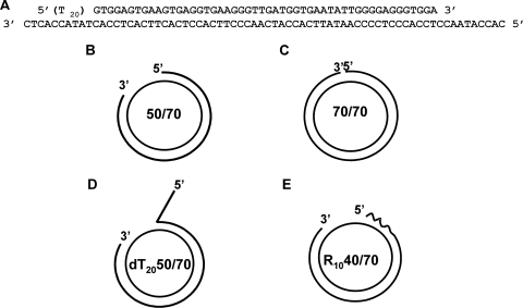
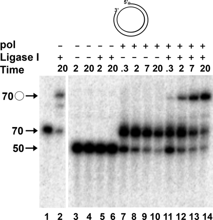
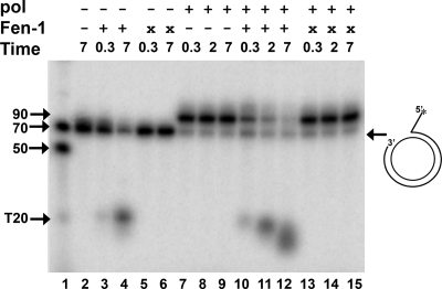
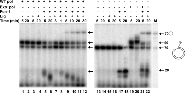
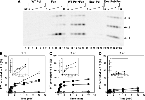
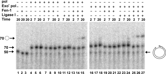
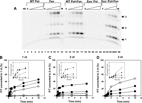
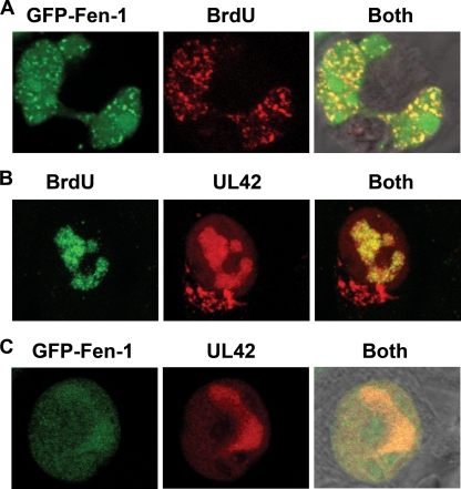
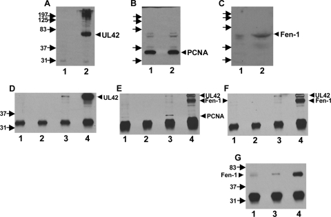
The processing of lagging-strand intermediates has not been demonstrated in vitro for herpes simplex virus type 1 (HSV-1). Human flap endonuclease-1 (Fen-1) was examined for its ability to produce ligatable products with model lagging-strand intermediates in the presence of the wild-type or exonuclease-deficient (exo(-)) HSV-1 DNA polymerase (pol). Primer/templates were composed of a minicircle single-stranded DNA template annealed to primers that contained 5' DNA flaps or 5' annealed DNA or RNA sequences. Gapped DNA primer/templates were extended but not significantly strand displaced by the wild-type HSV-1 pol, although significant strand displacement was observed with exo(-) HSV-1 pol. Nevertheless, the incubation of primer/templates containing 5' flaps with either wild-type or exo(-) HSV-1 pol and Fen-1 led to the efficient production of nicks that could be sealed with DNA ligase I. Both polymerases stimulated the nick translation activity of Fen-1 on DNA- or RNA-containing primer/templates, indicating that the activities were coordinated. Further evidence for Fen-1 involvement in HSV-1 DNA synthesis is suggested by the ability of a transiently expressed green fluorescent protein fusion with Fen-1 to accumulate in viral DNA replication compartments in infected cells and by the ability of endogenous Fen-1 to coimmunoprecipitate with an essential viral DNA replication protein in HSV-1-infected cells.


Figures

FIG. 1.
FIG. 1.
Model primer/templates. All templates employ an ssDNA circular molecule 70 nt in length annealed to a variety of primers. (A) The sequences common to all primer/templates (P/Ts) as well as the 20-nt oligo(dT) flap (T20) used for some of the P/Ts are shown. The bottom strand represents the circular DNA template that was prepared as described in Materials and Methods. The top strand represents the complementary 50-nt sequence in all primers and the 5′ T20 flap used for some. (B) The 50-nt DNA primer is annealed to the circular template to create a 20-nt gap. (C) The annealed DNA primer contains an additional 20 nt complementary to the circular template to create an open circular molecule with a ligatable nick. (D) The primer strand contains oligo(dT) residues at the 5′ end of the 50-nt complementary sequence to form a 5′ flap and a 20-nt gap at the 3′ end. (E) The 50-nt complementary primer is composed of 10 RNA residues at the 5′ end (wavy line) linked to 40 residues of DNA and annealed to the 70-nt circular template.
FIG. 2.
FIG. 2.
Gap filling by HSV-1 pol creates ligatable nicks. Control ligation reactions were performed by incubating 0.5 nM 70/70 primer/template (Fig. 1C) without DNA ligase I (lane 1) or with DNA ligase I (lane 2) for 20 min. In lanes 3 to 14, reactions were performed with 0.5 nM 50/70 P/T (Fig. 1B) in which the primer strand was 5′ end labeled. The DNA was preincubated in the absence (−) or presence (+) of 50 nM HSV-1 pol catalytic subunit (pol) and/or 30 nM human DNA ligase I for 5 min at 30°C in the presence of EDTA. Reactions were initiated by the addition of MgCl2 as described in Materials and Methods and were terminated at the times indicated with EDTA. Products were resolved by denaturing gel electrophoresis, and the amount of radioactivity in each was determined as a percentage of total radioactivity present as detailed in Materials and Methods. The migration positions of the 50-nt primer, the unligated 70-nt extended primer, and the 70-nt circular, ligated DNA (○) are shown.
FIG. 3.
FIG. 3.
Processing of model primer/templates by wild-type HSV-1 DNA polymerase and Fen-1 activities. The 5′-flapped model primer/template (dT2050/70), depicted in Fig. 1D and at the right in this figure, contained a 32P label at the 5′ end of the primer strand. All reaction mixes contained 0.5 nM annealed DNA, were preincubated in the presence of EDTA with the indicated enzymes for 5 min at 30°C, and were initiated by the addition of MgCl2 as described in Materials and Methods. Reactions were terminated by the addition of EDTA at the times (in min) indicated, the reaction mixes were boiled, and the products were analyzed by denaturing electrophoresis. The presence or absence of the indicated enzymes is designated by a plus and minus sign, respectively. In some cases, head-inactivated enzyme was added (×). The final concentration of active enzyme, when present, was 50 nM for pol and 0.01 U/μl for Fen-1. Reaction mixtures containing no enzyme (lane 2) were incubated continuously in the presence of EDTA and define the zero time point for all reactions. The migration positions of marker oligonucleotides, including a 50- and 70-nt sequence complementary to the minicircle and a 20-nt oligo(dT) (T20) (lane 1), are shown on the left. The migration position of the fully gap-filled product (90 nt) also is shown. The amount of radioactivity in each product was determined as a percentage of the total radioactivity present as detailed in Materials and Methods.
FIG. 4.
FIG. 4.
Flap cleavage and DNA ligation with HSV-1 wild-type and exonuclease-deficient polymerase. The dT2050/70 flapped primer/template (Fig. 1D and shown at the right in this figure) was used in all reactions, and the presence (+) or absence (−) of the wild-type HSV-1 pol catalytic subunit (WT pol; 50 nM), the exonuclease-deficient D368A pol catalytic subunit (Exo pol; 50 nM), Fen-1 (0.01 U/μl), and/or DNA ligase I (Lig; 30 nM) in reaction mixtures is indicated above each lane. To track all products, the primer strand was 5′ end labeled with 32P, and the dNTPs used for extension contained [α-32P]dATP. Reactions were performed at 30°C and terminated with EDTA at the indicated times (in minutes) following initiation with MgCl2. Products were resolved by denaturing electrophoresis through gels containing 12% polyacrylamide and 6 M urea. The migration positions of 20, 70, and 90 nt are shown, as is the circular product formed by the ligation of a control 70/70 P/T (lane M). Due to the presence of labeled dATP in reactions, the proportion of P/T extended was estimated from the radioactivity remaining at the 70-nt (linear) position compared to that present when no enzymes were added (lanes 13 and 14). The rate of the formation of ligated product was estimated by plotting the radioactivity in the bands corresponding to 70-nt circular product as a function of time and determining the slope of the linear portion by least-squares analysis.
FIG. 5.
FIG. 5.
Stimulation of human Fen-1 nick translation activity on DNA-containing primer/templates by HSV DNA polymerase. The 50/70 DNA-containing P/T was 5′ end labeled and used at a final concentration of 0.5 nM. (A) The P/T was incubated in the absence of enzyme (NE) but in the presence of EDTA (lane 1) or in the presence of 50 nM wild-type polymerase (WT Pol; lanes 2 to 6), Fen-1 (Fen) at a concentration of 0.01 U/μl (lanes 7 to 11), both enzymes together (WT Pol+Fen; lanes 13 to 17), 50 nM exo-deficient polymerase (Exo Pol; lanes 18 to 22), or exo-deficient pol together with Fen-1 (Exo- Pol+Fen; lanes 23 to 27). Reactions marked 0 (lanes 2, 7, 13, 18, and 23) contained the indicated enzymes but were incubated for 10 min in the presence of EDTA. Otherwise, reactions were terminated by the addition of EDTA at increasing times up to 10 min. Products were separated by electrophoresis through long, sequencing-style denaturing gels containing 20% polyacrylamide and 7 M urea. In this gel, only four time points are shown for each enzyme combination, corresponding to 20 s (lanes 3, 8, 14, 19, and 24), 40 s (lanes 4, 9, 15, 20, and 25), 2 min (lanes 5, 10, 16, 21, and 26), and 5 min (lanes 6, 11, 17, 22, and 27). Only the portion of the gel containing the mono- (1), di- (2), and trinucleotides (3) (arrows) is shown. The marker (M) (lanes 12 and 28) was produced by the partial digestion of the labeled single-stranded 50-nt DNA primer strand with 3′-to-5′ exo activity of HSV-1 pol in the absence of added nucleotides. (B, C, and D) The radioactivity of each of the small products (1, 2, and 3 nt, respectively) during the entire reaction period was calculated as a percentage of total radioactivity loaded onto each lane and plotted as a function of time (○, wild-type pol; □, exo-deficient pol; ⋄, Fen-1; •, wild-type pol + Fen-1; ▪, exo-deficient pol + Fen-1). The insets show the accumulation of products during the first 2 min. The initial rate of accumulation of each small product was estimated during the first 40 s from these data by least-squares analysis and is displayed in Table 1.
FIG. 6.
FIG. 6.
Processing of RNA-containing primer/templates by HSV-1 polymerase, Fen-1, and DNA ligase I. The R1040/70 P/T (Fig. 1E and depicted at the right in this figure) was used in all reactions and contained a 5′ end label on the primer. Reaction mixes included [α-32P]dATP to track all products. The migration positions of 50- and 70-nt linear DNA oligonucleotides and 70-nt circular molecules are shown with arrows. The presence (+) or absence (−) of the indicated enzymes is shown. Reactions were performed as indicated in the legend to Fig. 4 and separated by electrophoresis through 10% denaturing polyacrylamide gels. Product formation was estimated as described in the text.
FIG. 7.
FIG. 7.
RNA-containing P/T was incubated with wild-type or exo HSV-1 DNA polymerase and/or human Fen-1 as indicated. Reactions were performed as described in the legend to Fig. 5 and did not include any radioactively labeled dNTP, thus permitting the accurate quantification of all products. (A) Products were separated by electrophoresis through long, sequencing-style denaturing gels containing 20% polyacrylamide and 7 M urea together with DNA markers (lanes 12 and 28) prepared as described in the legend to Fig. 5. RNA oligonucleotides migrate slightly slower than DNA oligonucleotides, and the positions of these are shown by the arrows on the right. In this gel, only four time points are shown for each enzyme combination, corresponding to 20 s (lanes 3, 8, 14, 19, and 24), 40 s (lanes 4, 9, 15, 20, and 25), 2 min (lanes 5, 10, 16, 21, and 26), and 5 min (lanes 6, 11, 17, 22, and 27). Reactions marked 0 (lanes 2, 7, 13, 18, and 23) contained the indicated enzymes but were incubated for 10 min in the presence of EDTA. (B, C, and D) The radioactivity of each of the small products (1, 2, and 3 nt, respectively) during the entire reaction period was calculated as a percentage of total radioactivity loaded onto each lane and plotted as a function of time (○, wild-type pol; □, exo-deficient pol; ⋄, Fen-1; •, wild-type pol + Fen-1; ▪, exo-deficient pol + Fen-1). The insets show the accumulation of products during the first 2 min. The initial rate of accumulation of each small product was estimated during the first 40 s from these data by least-squares analysis and is displayed in Table 1.
FIG. 8.
FIG. 8.
Localization of Fen-1 and HSV-1 UL42 protein with replicating DNA in uninfected and infected cells. (A) BHK cells were transfected with 600 ng of plasmid expressing GFP-tagged Fen-1 (GFP-Fen-1). Thirty minutes prior to fixation (48 h posttransfection), BrdU was added to a final concentration of 100 μM to tag replicating DNA. Following fixation, cells were permeabilized with 0.25% Triton X-100 and incubated first with mouse monoclonal antibody to BrdU, followed by incubation with RITC-conjugated goat-anti-mouse antibody. Cells were viewed using a confocal microscope set to detect GFP-Fen-1 only as green color (left), BrdU only as red color (center), and the simultaneous localization of both (right). A multitrack mode was used to collect images sequentially. To ensure that there was no overlap in emission between channels, the green or red lasers were turned off sequentially prior to final image collection (data not shown). (B) BHK cells were infected with HSV-1 strain KOS at an input multiplicity of 5 PFU/cell and fixed at 3 h postinfection. Thirty minutes prior to fixation, BrdU was added to cultures. Cells were stained sequentially with BrdU mouse monoclonal antibody, FITC-conjugated goat anti-mouse IgG, primary polyclonal anti-peptide antibody UL42, the viral polymerase processivity factor, and secondary antibody RITC-conjugated goat anti-rabbit IgG. Images were collected as described in the text to detect the localization of BdrU in replicating DNA only (left) or HSV-1 UL42 only in infected cells (center). The colocalization of actively replicating DNA and viral replication compartments is shown by the yellow color (right). (C) BHK cells were transfected with plasmid (600 ng) expressing GFP-Fen-1 and infected 48 h later with HSV-1 (5 PFU/cell). Cells were fixed at 3 h postinfection, as indicated, and stained with rabbit antibody to UL42 and RITC-conjugated goat anti-rabbit IgG. Images were collected in multitrack mode to detect the localization of GFP-Fen-1 (left), UL42 (center), or both (right). A series of 20 Z-sections (0.35 μm thick) was obtained and used to create three-dimensional projections of the cells shown (Movie S1A to C).
FIG. 9.
FIG. 9.
Coimmunoprecipitation of Fen-1 with HSV-1 UL42. (A to C) Extracts of Vero cells that were mock infected (lane 1) or infected with HSV-1 at a multiplicity of infection of 10 PFU/cell and harvested at 8 h postinfection (lane 2) were prepared as described in Materials and Methods. The presence of UL42 (A), PCNA (B), and Fen-1 (C) in 10 μl extract (140 μg protein) was determined by immunoblotting using rabbit polyclonal anti-peptide antibody 834 directed to UL42 residues 360 to 377 (40), rabbit polyclonal antibody directed to a PCNA epitope mapping between residues 75 and 125, or rabbit polyclonal antibody directed to full-length human Fen-1. Primary antibodies were detected with horseradish peroxidase-conjugated goat-anti-rabbit IgG as described in Materials and Methods. The migration of molecular size standards (in kDa) is shown to the left. (D to G) Extracts from mock-infected (lane 1) or infected (lanes 2 to 4) cells were immunoprecipitated with monoclonal antibody to human Fen-1 (lanes 1 and 3), HSV-1 UL42 (lane 4), or the control human c-myc (lane 2). The immunoprecipitates were separated by SDS-PAGE and probed first for the presence of UL42 (D) and then stripped and probed successively with antibodies to Fen-1 (F) and PCNA (E) as described for panels A to C. In panel G, a portion of the immunoprecipitated material was separated on a distinct gel and probed with rabbit polyclonal antibody to Fen-1. There was an insufficient amount of the c-myc immunoprecipitate remaining for this purpose.

Similar articles

Cited by

References

    1. Ayyagari, R., X. V. Gomes, D. A. Gordenin, and P. M. J. Burgers. 2003. Okazaki fragment maturation in yeast. I. Distribution of functions between Fen1 and Dna2. J. Biol. Chem. 278:1618-1625. - PubMed
    1. Bae, S.-H., and Y.-S. Seo. 2000. Characterization of the enzymatic properties of the yeast Dna2 helicase/endonuclease suggests a new model for Okazaki fragment processing. J. Biol. Chem. 275:38022-38031. - PubMed
    1. Bentley, D. J., C. Harrison, A.-M. Ketchen, N. J. Redhead, K. Samuel, M. Waterfall, J. D. Ansell, and D. W. Melton. 2002. DNA ligase I null mouse cells show normal DNA repair activity but altered DNA replication and reduced genome stability. J. Cell Sci. 115:1551-1561. - PubMed
    1. Biswal, N., B. K. Murry, and M. Benyish-Melnick. 1974. Ribonucleotides in newly synthesized DNA of herpes simplex virus. Virology 69:87-99. - PubMed
    1. Boehmer, P. E., and A. V. Nimonkar. 2003. Herpes virus replication. IUBMB Life 55:13-22. - PubMed

Publication types