. 2010 Jul;84(14):6909-22.
doi: 10.1128/JVI.00081-10. Epub 2010 May 5.

Inefficient control of host gene expression by the 2009 pandemic H1N1 influenza A virus NS1 protein

Affiliations

Inefficient control of host gene expression by the 2009 pandemic H1N1 influenza A virus NS1 protein

Benjamin G Hale et al. J Virol. 2010 Jul.

Abstract

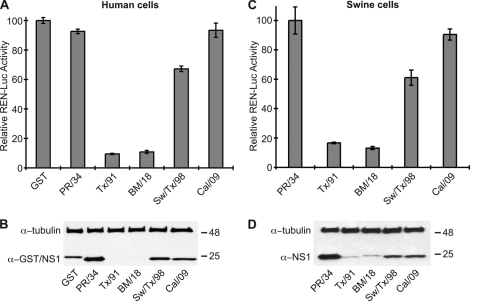
In 2009, a novel swine-origin H1N1 influenza A virus emerged. Here, we characterize the multifunctional NS1 protein of this human pandemic virus in order to understand factors that may contribute to replication efficiency or pathogenicity. Although the 2009 H1N1 virus NS1 protein (2009/NS1) is an effective interferon antagonist, we found that this NS1 (unlike those of previous human-adapted influenza A viruses) is unable to block general host gene expression in human or swine cells. This property could be restored in 2009/NS1 by replacing R108, E125, and G189 with residues corresponding to human virus consensus. Mechanistically, these previously undescribed mutations acted by increasing binding of 2009/NS1 to the cellular pre-mRNA processing protein CPSF30. A recombinant 2009 H1N1 influenza A virus (A/California/04/09) expressing NS1 with these gain-of-function substitutions was more efficient than the wild type at antagonizing host innate immune responses in primary human epithelial cells. However, such mutations had no significant effect on virus replication in either human or swine tissue culture substrates. Surprisingly, in a mouse model of pathogenicity, the mutant virus appeared to cause less morbidity, and was cleared faster, than the wild type. The mutant virus also demonstrated reduced titers in the upper respiratory tracts of ferrets; however, contact and aerosol transmissibility of the virus was unaffected. Our data highlight a potential human adaptation of NS1 that seems absent in "classically derived" swine-origin influenza A viruses, including the 2009 H1N1 virus. We discuss the impact that a natural future gain of this NS1 function may have on the new pandemic virus in humans.


Figures

FIG. 1.
FIG. 1.
Inhibition of IFN-β induction by different influenza A virus NS1 proteins. 293T cells were cotransfected for 16 h with a pCAGGS expression plasmid encoding the indicated NS1 protein (or GST) together with a FF-Luc IFN-β-promoter reporter plasmid (p125Luc). After infection with SeV for a further 12 h, FF-Luc activity was determined. The zoomed inset highlights differences between NS1 proteins. Results represent the means and standard deviations of triplicate values (normalized to GST plus SeV) obtained in a single experiment and are representative of results of two independent experiments.
FIG. 2.
FIG. 2.
The Cal/09 NS1 protein is unable to block general gene expression in human or swine cells. Human 293T (A) or swine PK-15 (C) cells were cotransfected with a pCAGGS expression plasmid encoding the indicated NS1 protein (or GST) together with a constitutively active Renilla luciferase plasmid. Luciferase activity was determined 24 h posttransfection. Results show the means and standard deviations of triplicate values (normalized to GST [A] or PR/34 NS1 [C]) obtained in a single experiment and are representative of results of two independent experiments. (B) Western blot analysis of lysates from panel A. (D) Western blot analysis of lysates from panel C. NS1 and GST were detected using a polyclonal rabbit anti-serum raised against a fusion protein of GST and the N-terminal RNA binding domain of NS1. Tubulin acted as a loading control. Molecular mass markers (in kilodaltons) are indicated to the right.
FIG. 3.
FIG. 3.
Residues at the interface of the NS1-CPSF30 complex. (A) Amino acid sequence alignment of the NS1 EDs (residues 85 to 203) from PR/34, BM/18, Tx/91, Sw/Tx/98, and Cal/09. Identity (*) and level of similarity (: or .) are indicated. Gray shading highlights residues of NS1 previously shown experimentally to be important for CPSF30 binding. Black boxes highlight residues common to PR/34, BM/18, and Tx/91 NS1 (human-like) EDs but different in Sw/Tx/98 and Cal/09 NS1 (swine-like) EDs. Residues 108, 125, and 189 (which are the only residues unique to Sw/Tx/98 and Cal/09 NS1 proteins at the NS1-CPSF30 interface; see below) are labeled. (B) Structural representation of NS1 in complex with the F2F3 fragment of CPSF30. The monomeric NS1 ED is shown in cartoon format (green), while a surface representation (blue) of the CPSF30-F2F3 fragment is shown. Unique residues of Sw/Tx/98 and Cal/09 NS1 EDs compared to PR/34, BM/18, and Tx/91 are highlighted with black spheres. (C) Close-up view of the NS1-CPSF30 interface with residues different in Sw/Tx/98 and Cal/09 NS1 EDs highlighted in black (sticks). Panels B and C were generated using MacPyMol (Protein Data Bank identification no. 2RHK [8]).
FIG. 4.
FIG. 4.
Three amino acid substitutions restore the ability of the Cal/09 NS1 protein to block general gene expression in human and swine cells. Human 293T (A) or swine PK-15 (C) cells were cotransfected with a pCAGGS expression plasmid encoding the indicated WT NS1 protein or the Cal/09 mutants, R108K, E125D, G189D, R108K/G189D (108/189), or R108K/E125D/G189D (TripleMut), together with a constitutively active Renilla luciferase plasmid. Luciferase activity was determined 24 h posttransfection. Results show the means and standard deviations of triplicate values (normalized to WT Cal/09 NS1) obtained in a single experiment and are representative of results of two independent experiments. (B and D) Western blot analysis of lysates from panels A and C, respectively. NS1 and tubulin were detected as described for Fig. 2.
FIG. 5.
FIG. 5.
Interaction of CPSF30 with WT Cal/09 NS1 or TripleMut Cal/09 NS1. FLAG-tagged CPSF30 was expressed in 293T cells (CPSF30), mixed with in vitro-synthesized [35S]methionine-labeled NS1 (WT Cal/09, TripleMut Cal/09, or HA-tagged Tx/91), and immunoprecipitated (IP) using anti-FLAG resin. 293T lysates without FLAG-tagged CPSF30 (Mock) acted as a negative control. Following SDS-PAGE, precipitated proteins were detected by Coomassie blue staining (IgG), Western blotting (FLAG-tagged CPSF30), or phosphorimaging ([35S]methionine-labeled NS1). Immunoprecipitations were performed using 500 mM NaCl (A, “In” denotes 10% input) or 200 mM NaCl (B, “In” denotes 1% input). (C) Ability of WT Cal/09 NS1 or TripleMut Cal/09 NS1 to bind synthetic dsRNA. Lysates from A549 cells infected with rCal/09 WT or rCal/09 TripleMut (MOI of 5 PFU/cell) were precipitated with pI:C-Sepharose (pI:C) or Sepharose only (−). Following SDS-PAGE, NS1 proteins were detected by Western blotting. Molecular mass markers (in kilodaltons) are indicated on the right.
FIG. 6.
FIG. 6.
In vitro characterization of the rCal/09 TripleMut virus. (A) Multicycle growth analysis of rCal/09 WT and nonrecombinant Cal/09 WT viruses in primary differentiated human airway epithelial cells. (B) Plaque phenotype of rCal/09 WT and rCal/09 TripleMut viruses in MDCK cells. (C) Western blot determination of viral NP and NS1 protein expression in MDCK cells infected for 12 h with the rCal/09 WT or rCal/09 TripleMut (TM) viruses (MOI of 5 PFU/cell). Molecular mass markers (in kilodaltons) are indicated to the right. Multicycle growth analysis of rCal/09 WT and rCal/09 TripleMut viruses in primary differentiated human airway epithelial cells (D) and the swine PK-15 cell line (E). Data points for growth curves show mean values of biological triplicates, and error bars represent standard deviations.
FIG. 7.
FIG. 7.
Ability of rCal/09 TripleMut virus to limit expression of IFN-β and IFN-stimulated genes during infection. Primary undifferentiated human airway epithelial cells were infected at an MOI of 5 PFU/cell with the rCal/09 WT or rCal/09 TripleMut virus. (A) Infectious titers determined by plaque assay of the supernatants after 24 h. (B to F) Cells were lysed at the times indicated, and total RNA was extracted. Following reverse transcription using oligo(dT), the levels of viral NA mRNA (B), IFN-β mRNA (C), OAS mRNA (D), MxA mRNA (E), and IP10 mRNA (F) were quantified in triplicate by qPCR. Values were averaged and normalized to 18S RNA. Mean induction levels relative to mRNA levels in mock-infected cells are shown. Error bars represent standard deviations. Means and standard deviations were calculated from biological triplicates.
FIG. 8.
FIG. 8.
Replication and pathogenicity of the rCal/09 TripleMut virus in mice. Seven-week-old DBA/2J mice were infected intranasally with 105 PFU of the rCal/09 WT or rCal/09 TripleMut virus. PBS-treated mice acted as a negative control. (A) Body weights were determined daily. Data show mean body weights of mice (n = 5). Error bars represent standard deviations. Significance was determined using two-tailed Student's t test (**, P < 0.01; *, P < 0.05). (B) Lung titers were determined on days 2, 4, and 7 postinfection from three DBA/2J mice infected in parallel with 104 PFU of the rCal/09 WT or rCal/09 TripleMut virus. Bars represent mean values. (C) Murine IFN-β mRNA was quantified from the lung homogenates used in panel B by qRT-PCR as described for Fig. 7.
FIG. 9.
FIG. 9.
Replication and transmission dynamics of the rCal/09 TripleMut virus in ferrets. Six- to 8-month-old female Fitch ferrets were infected intranasally with 106 TCID50 of the rCal/09 WT or rCal/09 TripleMut virus. (A) Viral titers in nasal turbinates, trachea, and lungs were determined on day 5 postinfection from two infected ferrets. Bars represent mean values. (B and C) Nasal wash titers for rCal/09 WT (B) and rCal/09 TripleMut (C) viruses were determined from ferrets infected intranasally (n = 2), by direct contact (n = 2), and by aerosol contact (large and small respiratory droplet) (n = 2) on the days indicated. Bars (dark and light gray) represent the raw results of two independent experiments. To limit the number of animals used, the rCal/09 WT-infected ferret data shown are the same as previously described (19). All experiments were performed in parallel to allow fair comparisons.

References

    1. Anonymous. Swine influenza A (H1N1) infection in two children—Southern California, March-April 2009. MMWR Morb. Mortal. Wkly. Rep. 58:400-402. - PubMed
    1. Babiuk, S., R. Albrecht, Y. Berhane, P. Marszal, J. A. Richt, A. Garcia-Sastre, J. Pasick, and H. Weingartl. 2010. 1918 and 2009 H1N1 influenza are not pathogenic in birds. J. Gen. Virol. 91:339-342. - PMC - PubMed
    1. Basler, C. F., A. H. Reid, J. K. Dybing, T. A. Janczewski, T. G. Fanning, H. Zheng, M. Salvatore, M. L. Perdue, D. E. Swayne, A. Garcia-Sastre, P. Palese, and J. K. Taubenberger. 2001. Sequence of the 1918 pandemic influenza virus nonstructural gene (NS) segment and characterization of recombinant viruses bearing the 1918 NS genes. Proc. Natl. Acad. Sci. U. S. A. 98:2746-2751. - PMC - PubMed
    1. Boon, A. C., J. deBeauchamp, A. Hollmann, J. Luke, M. Kotb, S. Rowe, D. Finkelstein, G. Neale, L. Lu, R. W. Williams, and R. J. Webby. 2009. Host genetic variation affects resistance to infection with a highly pathogenic H5N1 influenza A virus in mice. J. Virol. 83:10417-10426. - PMC - PubMed
    1. Brown, E. G., H. Liu, L. C. Kit, S. Baird, and M. Nesrallah. 2001. Pattern of mutation in the genome of influenza A virus on adaptation to increased virulence in the mouse lung: identification of functional themes. Proc. Natl. Acad. Sci. U. S. A. 98:6883-6888. - PMC - PubMed

Publication types

MeSH terms

Substances