. 2010 Jul;84(14):7243-55.
doi: 10.1128/JVI.02636-09. Epub 2010 May 5.

Ebola virus glycoprotein counteracts BST-2/Tetherin restriction in a sequence-independent manner that does not require tetherin surface removal

Affiliations

Ebola virus glycoprotein counteracts BST-2/Tetherin restriction in a sequence-independent manner that does not require tetherin surface removal

Lisa A Lopez et al. J Virol. 2010 Jul.

Abstract

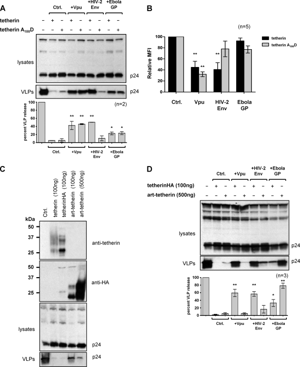
BST-2/tetherin is an interferon-inducible protein that restricts the release of enveloped viruses from the surface of infected cells by physically linking viral and cellular membranes. It is present at both the cell surface and in a perinuclear region, and viral anti-tetherin factors including HIV-1 Vpu and HIV-2 Env have been shown to decrease the cell surface population. To map the domains of human tetherin necessary for both virus restriction and sensitivity to viral anti-tetherin factors, we constructed a series of tetherin derivatives and assayed their activity. We found that the cytoplasmic tail (CT) and transmembrane (TM) domains of tetherin alone produced its characteristic cellular distribution, while the ectodomain of the protein, which includes a glycosylphosphatidylinositol (GPI) anchor, was sufficient to restrict virus release when presented by the CT/TM regions of a different type II membrane protein. To counteract tetherin restriction and remove it from the cell surface, HIV-1 Vpu required the specific sequence present in the TM domain of human tetherin. In contrast, the HIV-2 Env required only the ectodomain of the protein and was sensitive to a point mutation in this region. Strikingly, the anti-tetherin factor, Ebola virus GP, was able to overcome restriction conferred by both tetherin and a series of functional tetherin derivatives, including a wholly artificial tetherin molecule. Moreover, GP overcame restriction without significantly removing tetherin from the cell surface. These findings suggest that Ebola virus GP uses a novel mechanism to circumvent tetherin restriction.


Figures

FIG. 1.
FIG. 1.
Tetherin and derivatives. (A) Schematic of tetherin derivatives used in this study. Black represents tetherin sequences, gray represents TfR sequences, and the white octagon is EGFP. (B) Western blot of lysates of 293A cells transfected with 100 ng of the indicated tetherin plasmids or a control CMV expression plasmid (Ctrl.) and probed with anti-tetherin antibody. (C) FACS analysis of 293T cells transfected with 250 ng of a DsRed expression plasmid and 200 ng of the indicated tetherin plasmids (gray-shaded area) or control CMV plasmid (black line), stained with anti-tetherin antibody and gated on the DsRed-positive population. One representative data set, including peak fluorescence intensities, is shown from n = 3 independent experiments. (D) Restriction of HIV-1 VLP release from 293A cells cotransfected with 8 μg pHIV-1-pack and 100 ng of the indicated tetherin expression plasmids or CMV plasmid control (Ctrl.). Cell lysates and supernatants were analyzed by Western blotting with anti-p24 antibody, and the percent VLP release was calculated as the ratio of p24 signal in supernatants to lysates made relative to the control (no-tetherin) value. The graph shows the mean percent VLP release plus standard deviations for n = 3 experiments. All constructs except mini-tetherin significantly restricted virus release compared to the control. P < 0.01 (**).
FIG. 2.
FIG. 2.
Subcellular distribution of tetherin derivatives. (A) Western blot of HeLa cells transfected with 500 ng of the indicated tetherin plasmids and probed with anti-GFP antibody. (B) Ability of EGFP-tagged tetherin derivatives to restrict the release of HIV-1 VLPs from 293A cells, cotransfected with 8 μg pHIV-1-pack and 200 ng of the indicated tetherin plasmids. Cell lysates and pelleted supernatants were probed for p24 expression, and the percent VLP release was calculated. The graph shows means plus standard deviations for n = 2 experiments. Significance is indicated as P < 0.01 (**) compared to the control (no-tetherin) value. (C) Confocal analysis of endogenous tetherin present in fixed HeLa cells, detected with anti-tetherin antiserum for both nonpermeabilized and permeabilized cells. Scale bars represent 10 μm. Nonpermeabilized cells more clearly display the cell surface rim of tetherin. (D) Confocal analysis of EGFP-tagged tetherin and derivatives expressed in 293A and HeLa cells and detected using either anti-tetherin or anti-EGFP antibodies for nonpermeabilized and permeabilized cells, as indicated. Nuclei were stained with DAPI (blue). Scale bars represent 10 μm. All EGFP-tagged proteins were present at both the cell surface and in a perinuclear compartment.
FIG. 3.
FIG. 3.
Ability of viral anti-tetherin factors to overcome restriction by tetherin derivatives. 293A cells were transfected with 8 μg pHIV-1-pack, 100 ng of the indicated tetherin plasmids, and 2 μg of either a control CMV expression plasmid (Ctrl.) or expression plasmids for Vpu (pcDNA-Vphu), HIV-2 Env, or Ebola virus GP (pEboGP). VLP release was analyzed by the Western blotting of lysates and pelleted supernatants with anti-p24 antibody. Tetherin and viral proteins were detected using specific antisera. The extent of VLP release also was quantitated by the p24 ELISA of supernatants for n = 3 independent experiments and is shown as the mean percent (plus standard deviations) p24 in the supernatants relative to the amount produced in the absence of tetherin (Ctrl.). Statistical significance was determined by comparing levels of p24 in the presence of a viral anti-tetherin factor to the level achieved when the tetherin derivative alone was expressed and is indicated as P < 0.05 (*) or P < 0.01 (**).
FIG. 4.
FIG. 4.
Effect of Vpu on cell surface expression of tetherin derivates. (A) FACS analysis of 293T cells transfected with 200 ng of the indicated tetherin plasmids, 3 μg pcDNA-Vphu, and 250 ng of a DsRed expression plasmid. Tetherin derivatives were detected using anti-tetherin antiserum; mini-tetherin was detected using an anti-GFP antibody. One representative set of histograms from n = 3 independent experiments is shown. Solid black lines are tetherin levels when expressed alone, gray-shaded areas are tetherin levels in the presence of Vpu. (B) 293A cells were cotransfected with 100 ng of the indicated EGFP-tagged tetherin plasmids and 2 μg pVphu-HcRed, fixed, permeabilized, and stained with mouse anti-GFP antibody (green). Vpu-HcRed-expressing cells are shown as red, and nuclei are stained with DAPI (blue). Scale bars represent 10 μm.
FIG. 5.
FIG. 5.
Effect of HIV-2 Env on cell surface expression of tetherin derivates. (A) FACS analysis of 293T cells transfected with 200 ng of the indicated tetherin plasmids, 3 μg HIV-2Env plasmid, and 250 ng of a DsRed expression plasmid. Tetherin was detected using an anti-tetherin antiserum; mini-tetherin was detected using an anti-GFP antibody. One representative set of histograms from n = 3 independent experiments is shown. Solid black lines are tetherin levels when expressed alone, gray-shaded areas are tetherin levels in the presence of HIV-2 Env. (B) 293A cells were cotransfected with 100 ng of the indicated EGFP-tagged tetherin plasmids and 2 μg HIV-2 Env, fixed, permeabilized, and stained with mouse anti-GFP antibody (green), anti-HIV-2 Env antiserum (red), and DAPI (blue). Scale bars represent 10 μm.
FIG. 6.
FIG. 6.
Effect of Ebola virus GP on cell surface expression of tetherin derivates. (A) 293A cells were transfected with 8 μg of pHIV-1-pack, 70 ng of tetherin where indicated, and 2 μg of expression plasmids for either a control (Ctrl.) CMV plasmid or Ebola virus GP expression plasmid pEboGP or pEboGPiresDsRed. Lysates and supernatants were analyzed for p24 expression by Western blotting, and the percent VLP release was calculated. The graph shows means plus standard deviations for n = 3 independent experiments. Both Ebola GP plasmids increased the amount of VLP release compared to that of the tetherin-only control with P values of <0.05 (*) and <0.01 (**) as indicated, but they were not statistically different from each other in their ability to enhance VLP release. (B) 293A cells were cotransfected with the indicated tetherin plasmids and 2 μg pEboGPiresDsRed, fixed, permeabilized, and stained with mouse anti-GFP antibody (green). Ebola virus GP-expressing cells are red, and nuclei are stained with DAPI (blue). Scale bars represent 10 μm. (C) FACS analysis of 293T cells transfected with 200 ng of tetherin plasmids, 3 μg Ebola virus GP (pEboGP), and 250 ng of a DsRed expression plasmid. Tetherin was detected using an anti-tetherin antiserum. Solid black lines are tetherin levels when expressed alone, and the gray-shaded area indicates tetherin levels in the presence of Ebola virus GP. One representative set of histograms from n = 3 independent experiments is shown.
FIG. 7.
FIG. 7.
Activity of viral anti-tetherin factors against tetherinA100D and art-tetherin. (A) 293T cells were transfected with 8 μg of pHIV-1-pack, 100 ng of tetherin or tetherinA100D, and 2 μg of either a control (Ctrl.) CMV expression plasmid, Vpu (pcDNA-Vphu), HIV-2 Env, or Ebola virus GP (pEboGP). Lysates and supernatants were analyzed by Western blotting using anti-p24 antibody. One representative experiment from n = 2 is shown, together with a graphical representation of the mean percent VLP release plus standard deviations relative to results for the control (no tetherin). Statistical significance was determined by comparing VLP release in the presence of a viral anti-tetherin factor to the level achieved when either tetherin or tetherinA100D alone was expressed and is indicated as P < 0.05 (*) or P < 0.01 (**). (B) 293T cells were transfected with 200 ng of tetherin or tetherinA100D plasmids, 250 ng of a DsRed expression plasmid, and 3 μg Vpu (pcDNA-Vphu), HIV-2 Env, or Ebola virus GP (pEboGP) plasmid. Tetherin was detected using an anti-tetherin antiserum and FACS analysis, with viable cells gated on the DsRed-positive population, and acquired cells were analyzed for the mean fluorescence intensity (MFI) of tetherin expression. The graph shows means plus standard deviations for MFI in the presence of viral anti-tetherin factors relative to either the tetherin or tetherinA100D control for n = 5 independent experiments. Statistical significance was determined by comparing VLP release in the presence of a viral anti-tetherin factor to the level achieved with either tetherin or tetherinA100D alone and is indicated as P < 0.01 (**). (C) 293T cells were cotransfected with 8 μg pHIV-1-pack and the indicated amounts of either tetherin, tetherinHA, or art-tetherin. Cell lysates and VLPs pelleted from supernatants were analyzed by Western blotting. (D) 293T cells were transfected with 8 μg pHIV-1-pack, 100 ng of either tetherinHA or 500 ng of art-tetherin plasmid, and 2 μg of the indicated expression plasmids. Lysates and supernatants were analyzed by Western blotting using anti-p24 antibody. The graph shows means plus standard deviations for n = 3 independent experiments. Statistical significance was determined by comparing VLP release in the presence of a viral anti-tetherin factor to the level achieved with either tetherinHA or art-tetherin alone and is indicated as P < 0.05 (*) or P < 0.01 (**).

Similar articles

Cited by

References

    1. Abada, P., B. Noble, and P. M. Cannon. 2005. Functional domains within the human immunodeficiency virus type 2 envelope protein required to enhance virus production. J. Virol. 79:3627-3638. - PMC - PubMed
    1. Andrew, A. J., E. Miyagi, S. Kao, and K. Strebel. 2009. The formation of cysteine-linked dimers of BST-2/tetherin is important for inhibition of HIV-1 virus release but not for sensitivity to Vpu. Retrovirology 6:80. - PMC - PubMed
    1. Bartee, E., A. McCormack, and K. Fruh. 2006. Quantitative membrane proteomics reveals new cellular targets of viral immune modulators. PLoS Pathog. 2:e107. - PMC - PubMed
    1. Bour, S., H. Akari, E. Miyagi, and K. Strebel. 2003. Naturally occurring amino acid substitutions in the HIV-2 ROD envelope glycoprotein regulate its ability to augment viral particle release. Virology 309:85-98. - PubMed
    1. Douglas, J. L., K. Viswanathan, M. N. McCarroll, J. K. Gustin, K. Fruh, and A. V. Moses. 2009. Vpu directs the degradation of the human immunodeficiency virus restriction factor BST-2/Tetherin via a βTrCP-dependent mechanism. J. Virol. 83:7931-7947. - PMC - PubMed

Publication types

MeSH terms

Substances