. 2010 Jul;84(14):7312-24.
doi: 10.1128/JVI.00209-10. Epub 2010 May 5.

Vif of feline immunodeficiency virus from domestic cats protects against APOBEC3 restriction factors from many felids

Affiliations

Vif of feline immunodeficiency virus from domestic cats protects against APOBEC3 restriction factors from many felids

Jörg Zielonka et al. J Virol. 2010 Jul.

Abstract

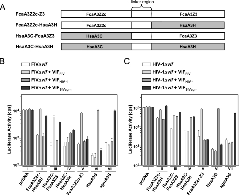
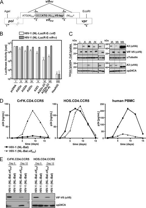
To get more insight into the role of APOBEC3 (A3) cytidine deaminases in the species-specific restriction of feline immunodeficiency virus (FIV) of the domestic cat, we tested the A3 proteins present in big cats (puma, lion, tiger, and lynx). These A3 proteins were analyzed for expression and sensitivity to the Vif protein of FIV. While A3Z3s and A3Z2-Z3s inhibited Deltavif FIV, felid A3Z2s did not show any antiviral activity against Deltavif FIV or wild-type (wt) FIV. All felid A3Z3s and A3Z2-Z3s were sensitive to Vif of the domestic cat FIV. Vif also induced depletion of felid A3Z2s. Tiger A3s showed a moderate degree of resistance against the Vif-mediated counter defense. These findings may imply that the A3 restriction system does not play a major role to prevent domestic cat FIV transmission to other Felidae. In contrast to the sensitive felid A3s, many nonfelid A3s actively restricted wt FIV replication. To test whether Vif(FIV) can protect also the distantly related human immunodeficiency virus type 1 (HIV-1), a chimeric HIV-1.Vif(FIV) was constructed. This HIV-1.Vif(FIV) was replication competent in nonpermissive feline cells expressing human CD4/CCR5 that did not support the replication of wt HIV-1. We conclude that the replication of HIV-1 in some feline cells is inhibited only by feline A3 restriction factors and the absence of the appropriate receptor or coreceptor.


Figures

FIG. 1.
FIG. 1.
Representation of APOBEC3 (A3) coding regions in the genome of Felis catus. (A) Transcripts with translated exons (Z2, gray rectangles; Z3, black rectangles) and spliced-out introns (dashed lines) are indicated. Three readthrough transcripts were found: the already described two-domain FcaA3Z2c-Z3 (31) and the two splice variants (SV) A3Z2b-Z3 (SV-A) and A3Z2b-Z3 (SV-B) that include the exon 4-derived sequence of either A3Z2b or -Z2c in an additionally spliced version (for details, see reference 31). (B) Schematic representation of the exon structure of the A3Z2-Z3 readthrough transcripts. The locations of amino acid replacements in FcaA3Z2c-Z3 are indicated with respect to exon 4 variability of major cat breeds (upper figure) and A3Z2-Z3 genes of big cats (lower figure). A plus sign indicates the insertion of an amino acid. Numbers in shaded boxes indicate exons from Z2c, and those in open boxes indicate exons from Z3. The region of Z2-Z3 proteins designated “linker region” protein is encoded by exon 2 of A3Z3, a sequence that is untranslated in single-domain A3Z3. BIR, Birman; BOB, Japanese Bobtail; SHO, British Shorthair; VAN, Turkish Van; Pti, Panthera tigris corbetti; Ppa, Panthera pardus japonensis; Ple, Panthera leo bleyenberghi; Lly, Lynx lynx; Pco, Puma concolor.
FIG. 2.
FIG. 2.
FIV Vif of domestic cats overcomes the antiviral activity of Felidae A3s. VSV-G pseudotyped wt FIV (expressing Vif), Δvif, and Δvif plus VifFIV luciferase reporter vectors were produced in 293T cells cotransfected with HA-tagged A3 expression plasmids of the domestic cat and chimeric A3s with major amino acid exchanges of exon 4 of A3Z2c of four cat breeding lines (A and B) and big cats (C and D). Roman numbers indicate a set of transfections with the same A3 plasmid. Plasmid pVifFIV expresses a V5-tagged Vif protein. pcDNA3.1(+) (pcDNA) was included as a control. Reporter vector infectivity was determined by quantification of luciferase activity in HOS cells transduced with vector particles normalized for RT. Asterisks represent statistically significant differences (*, P < 0.05; **, P < 0.01 [Dunnett t test]) relative to the pcDNA control. A3 and Vif (Vif-V5) expression in the transfected 293T producer cells were detected by immunoblotting with anti-HA antibody for A3 or anti-V5 for Vif (B and D). Cell lysates were also analyzed for equal amounts of total proteins by using anti-tubulin antibody. For panel B, Felis catus A3s proteins were also analyzed for encapsidation into released FIV particles. Encapsidated A3 proteins were detected on immunoblots probed with anti-HA antibody. Vector particle lysates were analyzed for equal amounts of viral proteins by using anti-VSV-G antibody. (E) Increasing amounts of A3 (0, 4, 20, 100, and 500 ng of plasmid) were tested against Δvif FIV and Δvif FIV plus Vif (500 ng of pVifFIV plasmid) and alternatively Δvif FIV in the presence of 500 ng of A3 plasmid with increasing amounts of VifFIV (0, 4, 20, 100, and 500 ng of plasmid) was analyzed (F). Corresponding immunoblots of lysates of the vector producer cells are shown. A3 and Vif (Vif-V5) expression in transfected 293T producer cells were detected by immunoblotting with anti-HA antibody for A3 or anti-V5 for Vif. Cell lysates were also analyzed for equal amounts of total proteins by using anti-tubulin antibody. α, anti; cps, counts per second; BIR, Birman; BOB, Japanese Bobtail; SHO, British Shorthair; VAN, Turkish Van; Fca, Felis catus; Pti, Panthera tigris corbetti; Ppa, Panthera pardus japonensis; Ple, Panthera leo bleyenberghi; Lly, Lynx lynx; Pco, Puma concolor; Hsa, Homo sapiens; (var1), A3Z2 variant transcript detected in the indicated species.
FIG. 2.
FIG. 2.
FIV Vif of domestic cats overcomes the antiviral activity of Felidae A3s. VSV-G pseudotyped wt FIV (expressing Vif), Δvif, and Δvif plus VifFIV luciferase reporter vectors were produced in 293T cells cotransfected with HA-tagged A3 expression plasmids of the domestic cat and chimeric A3s with major amino acid exchanges of exon 4 of A3Z2c of four cat breeding lines (A and B) and big cats (C and D). Roman numbers indicate a set of transfections with the same A3 plasmid. Plasmid pVifFIV expresses a V5-tagged Vif protein. pcDNA3.1(+) (pcDNA) was included as a control. Reporter vector infectivity was determined by quantification of luciferase activity in HOS cells transduced with vector particles normalized for RT. Asterisks represent statistically significant differences (*, P < 0.05; **, P < 0.01 [Dunnett t test]) relative to the pcDNA control. A3 and Vif (Vif-V5) expression in the transfected 293T producer cells were detected by immunoblotting with anti-HA antibody for A3 or anti-V5 for Vif (B and D). Cell lysates were also analyzed for equal amounts of total proteins by using anti-tubulin antibody. For panel B, Felis catus A3s proteins were also analyzed for encapsidation into released FIV particles. Encapsidated A3 proteins were detected on immunoblots probed with anti-HA antibody. Vector particle lysates were analyzed for equal amounts of viral proteins by using anti-VSV-G antibody. (E) Increasing amounts of A3 (0, 4, 20, 100, and 500 ng of plasmid) were tested against Δvif FIV and Δvif FIV plus Vif (500 ng of pVifFIV plasmid) and alternatively Δvif FIV in the presence of 500 ng of A3 plasmid with increasing amounts of VifFIV (0, 4, 20, 100, and 500 ng of plasmid) was analyzed (F). Corresponding immunoblots of lysates of the vector producer cells are shown. A3 and Vif (Vif-V5) expression in transfected 293T producer cells were detected by immunoblotting with anti-HA antibody for A3 or anti-V5 for Vif. Cell lysates were also analyzed for equal amounts of total proteins by using anti-tubulin antibody. α, anti; cps, counts per second; BIR, Birman; BOB, Japanese Bobtail; SHO, British Shorthair; VAN, Turkish Van; Fca, Felis catus; Pti, Panthera tigris corbetti; Ppa, Panthera pardus japonensis; Ple, Panthera leo bleyenberghi; Lly, Lynx lynx; Pco, Puma concolor; Hsa, Homo sapiens; (var1), A3Z2 variant transcript detected in the indicated species.
FIG. 3.
FIG. 3.
Inhibition of FIV by nonfeline A3s. VSV-G pseudotyped FIV luciferase reporter vectors (wt FIV, expressing Vif; Δvif; and Δvif plus VifFIV) were produced in 293T cells in the presence or absence (pcDNA) of human A3s (A) or nonprimate equine, murine, and porcine A3s (B). The infectivity of the vector particles was determined by quantification of luciferase activity in HOS cells transduced with equal amounts of particles. pcDNA, pcDNA3.1(+); SV, splice variant; hapI, haplotype I; hapII, haplotype II; cps, counts per second; Eca, Equus caballus; Mmus, Mus musculus; Ssc, Sus scrofa. Roman numerals indicate a set of transfections with the same A3 plasmid.
FIG. 4.
FIG. 4.
Analyses of human/feline A3 chimeras reveal Vif interactions with both Z-domains of FcaA3Z2c-Z3. (A) Schematic representation of human/feline A3Z2-Z3 chimera. Open bars denote feline sequences; filled bars denote human sequences. In FcaZ2-Z3, the Z2 and Z3 domains are naturally separated by a linker region (demarcated by dotted lines) that is encoded by exon 2 of Z3, which is untranslated in FcaA3Z3 (see Fig. 1A). FIVΔvif (B) and HIVΔvif (C) luciferase reporter particles were produced in 293T cells in the presence or absence (pcDNA) of feline and chimeric human/feline A3Z2-Z3s expression plasmids and expression plasmids for VifFIV, VifHIV-1, or VifSIVagm. To control the specificity of primate Vifs, human and AGM A3G expression plasmids were included. The infectivities of the vector particles were determined by quantification of luciferase in HOS cells transduced with equal amounts of vector particles. pcDNA, pcDNA3.1(+); cps, counts per second. Roman numerals indicate a set of transfections with the same A3 plasmid.
FIG. 5.
FIG. 5.
HIV-1 is protected by VifFIV in feline cells. (A) Schematic representation of the insertion of the vifFIV (shaded) in the vif gene of the HIV-derived constructs (NL-LucREvifFIV and NL-Bal.vifFIV) by fusion PCR. Overlapping pol (schematically depicted) and vpr genes are shown. Restriction enzyme recognition sites AgeI and EcoRI were used to clone the fusion PCR product. Position of PCR primers used are shown as arrows: primer 1, vpu_mut_5′out_HH; primer 2, HIV1_vif_FIV1.rv; primer 3, HIV1_vif_FIV2.fw; primer 4, HIV1_vif_FIV2a.rv; primer 5, HIV1_vif_FIV3.fw; and primer 6, HIV1_vif_FIV3.rv. “N” represents adenine (A), cytosine (C), guanine (G), or thymidine (T). (N)number represents the number of Ns. *, Stop codon. (B and C) The effect of VifFIV in cis for the infectivity of the chimeric HIV-1 constructs was tested by using single-cycle luciferase reporter viruses. VSV-G pseudotyped virus was produced by cotransfection of pNL-LucREΔvif or pNL-LucREvifFIV in the presence or absence (pcDNA) of the indicated expression plasmids for feline A3 and human A3G. Infectivity of the viruses was determined by quantification of luciferase in HOS cells infected with normalized amounts of viruses (B). Asterisks represent statistically significant differences (*, P < 0.05; **, P < 0.01 [Dunnett t test]) relative to the pcDNA control. Corresponding immunoblots of cell lysates and virion lysates were probed with anti-HA antibody for A3 expression (C). The expression of VifFIV was confirmed by probing the blot with an anti-V5 antibody. Cell lysates were also analyzed for equal amounts of total proteins by using an anti-tubulin antibody. Virus lysates were analyzed for equal amounts of viral proteins by using anti-p24CA (HIV-1 capsid) antibody. Roman numbers indicate a set of transfections with the same A3 plasmid. (D and E) Replication of wild-type HIV-1(NL-Bal) and HIV-1(NL-Bal.vifFIV). Human HOS.CD4.CCR5 cells, feline CrFK.CD4.CCR5 cells, and PHA/IL-2 activated human PBMC were infected with NL-BaL and NL-BaL.vifFIV at a multiplicity of infection of 0.05. (D) Culture supernatants were quantified every 2 or 3 days by p24CA ELISA. (E) Immunoblot analysis of virus lysates from infected CrFK.CD4.CCR5 and HOS.CD4.CCR5 cells on days 5 and 12 postinfection. Expression of VifFIV was confirmed by probing the filter with an anti-V5 antibody, the same blot was also stained with anti-p24CA (capsid) antibody to detect HIV-1. α, anti.

Similar articles

Cited by

References

    1. An, P., G. Bleiber, P. Duggal, G. Nelson, M. May, B. Mangeat, I. Alobwede, D. Trono, D. Vlahov, S. Donfield, J. J. Goedert, J. Phair, S. Buchbinder, S. J. O'Brien, A. Telenti, and C. A. Winkler. 2004. APOBEC3G genetic variants and their influence on the progression to AIDS. J. Virol. 78:11070-11076. - PMC - PubMed
    1. Bishop, K. N., R. K. Holmes, and M. H. Malim. 2006. Antiviral potency of APOBEC proteins does not correlate with cytidine deamination. J. Virol. 80:8450-8458. - PMC - PubMed
    1. Bogerd, H. P., B. P. Doehle, H. L. Wiegand, and B. R. Cullen. 2004. A single amino acid difference in the host APOBEC3G protein controls the primate species specificity of HIV type 1 virion infectivity factor. Proc. Natl. Acad. Sci. U. S. A. 101:3770-3774. - PMC - PubMed
    1. Bogerd, H. P., R. L. Tallmadge, J. L. Oaks, S. Carpenter, and B. R. Cullen. 2008. Equine infectious anemia virus resists the antiretroviral activity of equine APOBEC3 proteins through a packaging-independent mechanism. J. Virol. 82:11889-11901. - PMC - PubMed
    1. Carpenter, M. A., E. W. Brown, M. Culver, W. E. Johnson, J. Pecon-Slattery, D. Brousset, and S. J. O'Brien. 1996. Genetic and phylogenetic divergence of feline immunodeficiency virus in the puma (Puma concolor). J. Virol. 70:6682-6693. - PMC - PubMed

Publication types

MeSH terms