Evidence for DNA hairpin recognition by Zta at the Epstein-Barr virus origin of lytic replication
- PMID: 20444899
- PMCID: PMC2898250
- DOI: 10.1128/JVI.02666-09
Evidence for DNA hairpin recognition by Zta at the Epstein-Barr virus origin of lytic replication
Abstract
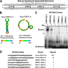
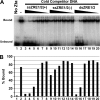
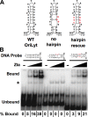
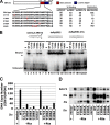
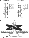
The Epstein-Barr virus immediate-early protein (Zta) plays an essential role in viral lytic activation and pathogenesis. Zta is a basic zipper (b-Zip) domain-containing protein that binds multiple sites in the viral origin of lytic replication (OriLyt) and is required for lytic-cycle DNA replication. We present evidence that Zta binds to a sequence-specific, imperfect DNA hairpin formed by an inverted repeat within the upstream essential element (UEE) of OriLyt. Mutations in the OriLyt sequence that are predicted to disrupt hairpin formation also disrupt Zta binding in vitro. Restoration of the hairpin rescues the defect. We also show that OriLyt DNA isolated from replicating cells contains a nuclease-sensitive region that overlaps with the inverted-repeat region of the UEE. Furthermore, point mutations in Zta that disrupt specific recognition of the UEE hairpin are defective for activation of lytic replication. These data suggest that Zta acts by inducing and/or stabilizing a DNA hairpin structure during productive infection. The DNA hairpin at OriLyt with which Zta interacts resembles DNA structures formed at other herpesvirus origins and may therefore represent a common secondary structure used by all herpesvirus family members during the initiation of DNA replication.
Figures


Similar articles
-
A redox-sensitive cysteine in Zta is required for Epstein-Barr virus lytic cycle DNA replication.J Virol. 2005 Nov;79(21):13298-309. doi: 10.1128/JVI.79.21.13298-13309.2005. J Virol. 2005. PMID: 16227252 Free PMC article.
-
The Epstein-Barr virus lytic transactivator Zta interacts with the helicase-primase replication proteins.J Virol. 1998 Nov;72(11):8559-67. doi: 10.1128/JVI.72.11.8559-8567.1998. J Virol. 1998. PMID: 9765394 Free PMC article.
-
Multivalent sequence recognition by Epstein-Barr virus Zta requires cysteine 171 and an extension of the canonical B-ZIP domain.J Virol. 2006 Nov;80(22):10942-9. doi: 10.1128/JVI.00907-06. Epub 2006 Sep 13. J Virol. 2006. PMID: 16971443 Free PMC article.
-
Regulation of cellular and viral protein expression by the Epstein-Barr virus transcriptional regulator Zta: implications for therapy of EBV associated tumors.Cancer Biol Ther. 2009 Jun;8(11):987-95. doi: 10.4161/cbt.8.11.8369. Epub 2009 Jun 10. Cancer Biol Ther. 2009. PMID: 19448399 Review.
-
Latent and lytic Epstein-Barr virus replication strategies.Rev Med Virol. 2005 Jan-Feb;15(1):3-15. doi: 10.1002/rmv.441. Rev Med Virol. 2005. PMID: 15386591 Review.
Cited by
-
Epstein-Barr virus lytic reactivation regulation and its pathogenic role in carcinogenesis.Int J Biol Sci. 2016 Oct 18;12(11):1309-1318. doi: 10.7150/ijbs.16564. eCollection 2016. Int J Biol Sci. 2016. PMID: 27877083 Free PMC article. Review.
-
Replication of Epstein-Barr viral DNA.Cold Spring Harb Perspect Biol. 2013 Jan 1;5(1):a013029. doi: 10.1101/cshperspect.a013029. Cold Spring Harb Perspect Biol. 2013. PMID: 23284049 Free PMC article. Review.
-
Sequencing of bovine herpesvirus 4 v.test strain reveals important genome features.Virol J. 2011 Aug 16;8:406. doi: 10.1186/1743-422X-8-406. Virol J. 2011. PMID: 21846388 Free PMC article.
-
Initiation of Epstein-Barr virus lytic replication requires transcription and the formation of a stable RNA-DNA hybrid molecule at OriLyt.J Virol. 2011 Mar;85(6):2837-50. doi: 10.1128/JVI.02175-10. Epub 2010 Dec 29. J Virol. 2011. PMID: 21191028 Free PMC article.
-
()-Epigallocatechin-3-Gallate Inhibits EBV Lytic Replication via Targeting LMP1-Mediated MAPK Signal Axes.Oncol Res. 2021 Sep 7;28(7):763-778. doi: 10.3727/096504021X16135618512563. Epub 2021 Feb 17. Oncol Res. 2021. PMID: 33629943 Free PMC article.
References
-
- Andersson, J. 2000. An overview of Epstein-Barr virus: from discovery to future directions for treatment and prevention. Herpes 7:76-82. - PubMed
-
- Arbuckle, M. I., and N. D. Stow. 1993. A mutational analysis of the DNA-binding domain of the herpes simplex virus type 1 UL9 protein. J. Gen. Virol. 74(Pt. 7):1349-1355. - PubMed
-
- Aslani, A., M. Olsson, and P. Elias. 2002. ATP-dependent unwinding of a minimal origin of DNA replication by the origin-binding protein and the single-strand DNA-binding protein ICP8 from herpes simplex virus type I. J. Biol. Chem. 277:41204-41212. - PubMed
-
- Aslani, A., S. Simonsson, and P. Elias. 2000. A novel conformation of the herpes simplex virus origin of DNA replication recognized by the origin binding protein. J. Biol. Chem. 275:5880-5887. - PubMed
Publication types
MeSH terms
Substances
Grants and funding
LinkOut - more resources
Full Text Sources

