

Deletion of androgen receptor in the smooth muscle of the seminal vesicles impairs secretory function and alters its responsiveness to exogenous testosterone and estradiol
- PMID: 20444943
- PMCID: PMC3033689
- DOI: 10.1210/en.2009-1339
Deletion of androgen receptor in the smooth muscle of the seminal vesicles impairs secretory function and alters its responsiveness to exogenous testosterone and estradiol
Abstract
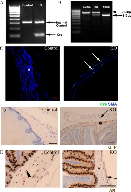
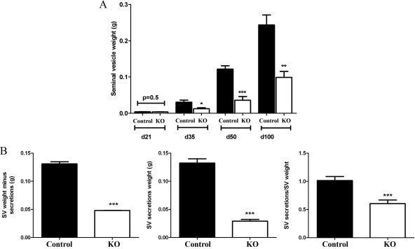
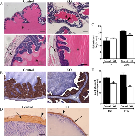
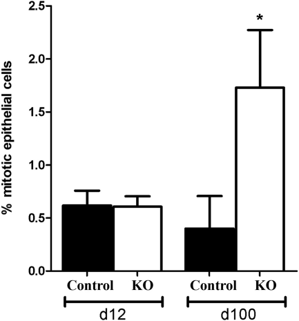
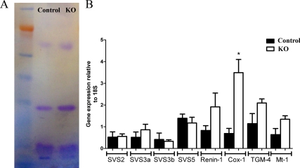
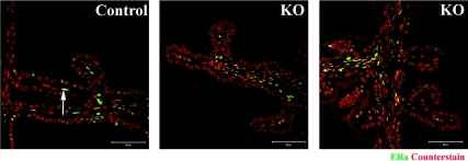
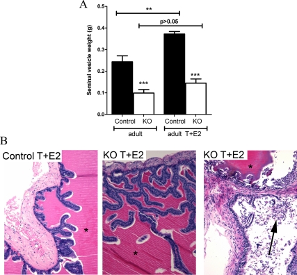
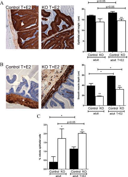
The seminal vesicles (SVs), like much of the male reproductive tract, depend on androgen-driven stromal-epithelial interactions for normal development, structure, and function. The primary function of the SVs is to synthesize proteins that contribute to the seminal plasma and this is androgen dependent. However, the cell-specific role for androgen action in adult SVs remains unclear. This study analyzed the SV in mice with targeted ablation of androgen receptors specifically in smooth muscle cells (PTM-ARKO) to determine in vivo whether it is androgen action in a subset of the SV stroma, the smooth muscle cells, that drives epithelial function and identity. These mice have significantly smaller SVs in adulthood with less smooth muscle and reduced epithelial cell height. Less epithelial cell proliferation was observed in adult PTM-ARKO SVs, compared with controls, and production of seminal proteins was reduced, indicating global impairment of epithelial cell function in PTM-ARKO SVs. None of these changes could be explained by altered serum testosterone or estradiol concentrations. We also demonstrate altered SV responsiveness to exogenous testosterone and estradiol in PTM-ARKO mice, indicating that smooth muscle androgen receptors may limit the SV epithelial proliferative response to exogenous estrogens. These results therefore demonstrate that the smooth muscle cells play a vital role in androgen-driven stromal-epithelial interactions in the SV, determining epithelial cell structure and function as well as limiting the SV epithelial proliferative response to exogenous estrogens.
Figures
Similar articles
-
Altered prostate epithelial development and IGF-1 signal in mice lacking the androgen receptor in stromal smooth muscle cells.Prostate. 2011 Apr;71(5):517-24. doi: 10.1002/pros.21264. Epub 2010 Oct 13. Prostate. 2011. PMID: 20945497 Free PMC article.
-
Direct response of the murine prostate gland and seminal vesicles to estradiol.Endocrinology. 2002 Dec;143(12):4922-33. doi: 10.1210/en.2002-220493. Endocrinology. 2002. PMID: 12446620
-
Morphogenetic and proliferative effects of testosterone and insulin on the neonatal mouse seminal vesicle in vitro.Endocrinology. 1991 Nov;129(5):2289-97. doi: 10.1210/endo-129-5-2289. Endocrinology. 1991. PMID: 1935767
-
Mucosa-Dependent, Stretch-Sensitive Spontaneous Activity in Seminal Vesicle.Adv Exp Med Biol. 2019;1124:217-231. doi: 10.1007/978-981-13-5895-1_9. Adv Exp Med Biol. 2019. PMID: 31183829 Review.
-
cis-elements required for expression of human protein C inhibitor gene in HepG2 cells and its androgen-dependent expression in rat reproductive organs.Semin Thromb Hemost. 2000;26(1):75-83. doi: 10.1055/s-2000-9807. Semin Thromb Hemost. 2000. PMID: 10805286 Review.
Cited by
-
Androgen receptor (AR) physiological roles in male and female reproductive systems: lessons learned from AR-knockout mice lacking AR in selective cells.Biol Reprod. 2013 Jul 25;89(1):21. doi: 10.1095/biolreprod.113.109132. Print 2013 Jul. Biol Reprod. 2013. PMID: 23782840 Free PMC article. Review.
-
Spongian diterpenoids inhibit androgen receptor activity.Mol Cancer Ther. 2013 May;12(5):621-31. doi: 10.1158/1535-7163.MCT-12-0978. Epub 2013 Feb 26. Mol Cancer Ther. 2013. PMID: 23443807 Free PMC article.
-
Autocrine androgen action is essential for Leydig cell maturation and function, and protects against late-onset Leydig cell apoptosis in both mice and men.FASEB J. 2015 Mar;29(3):894-910. doi: 10.1096/fj.14-255729. Epub 2014 Nov 17. FASEB J. 2015. PMID: 25404712 Free PMC article.
-
Ablation of the androgen receptor from vascular smooth muscle cells demonstrates a role for testosterone in vascular calcification.Sci Rep. 2016 Apr 20;6:24807. doi: 10.1038/srep24807. Sci Rep. 2016. PMID: 27095121 Free PMC article.
-
Transcriptomic analysis of the seminal vesicle response to the reproductive toxicant acrylamide.BMC Genomics. 2021 Oct 8;22(1):728. doi: 10.1186/s12864-021-07951-1. BMC Genomics. 2021. PMID: 34625024 Free PMC article.
References
-
- George FW, Wilson J 1994 Gonads and ducts in mammals. In: Knobil E, Neill JD, eds. The physiology of reproduction. 2nd ed. New York: Raven Press; 3–27
-
- Mooradian AD, Morley JE, Korenman SG 1987 Biological actions of androgens. Endocr Rev 8:1–28 - PubMed
-
- Wilson JD, George FW, Griffin JE 1981 The hormonal control of sexual development. Science 211:1278–1284 - PubMed
-
- Pointis G, Latreille MT, Cedard L 1980 Gonado-pituitary relationships in the fetal mouse at various times during sexual differentiation. J Endocrinol 86:483–488 - PubMed
-
- Quigley CA, De Bellis A, Marschke KB, el-Awady MK, Wilson EM, French FS 1995 Androgen receptor defects: historical, clinical, and molecular perspectives. Endocr Rev 16:271–321 - PubMed
Publication types
MeSH terms
Substances
Grants and funding
- MC_U127685841/MRC_/Medical Research Council/United Kingdom
- MC_U127685844/MRC_/Medical Research Council/United Kingdom
- MC_U127684422/MRC_/Medical Research Council/United Kingdom
- MC_U127685843/MRC_/Medical Research Council/United Kingdom
- U.1276.00.002.00003.01 (85841)/MRC_/Medical Research Council/United Kingdom
LinkOut - more resources
Full Text Sources
Molecular Biology Databases
