. 2010 Jul;151(7):3407-19.
doi: 10.1210/en.2009-1321. Epub 2010 May 5.

Interaction of adenosine 3',5'-cyclic monophosphate and tumor necrosis factor-alpha on serum amyloid A3 expression in mouse granulosa cells: dependence on CCAAT-enhancing binding protein-beta isoform

Affiliations

Interaction of adenosine 3',5'-cyclic monophosphate and tumor necrosis factor-alpha on serum amyloid A3 expression in mouse granulosa cells: dependence on CCAAT-enhancing binding protein-beta isoform

Deok-Soo Son et al. Endocrinology. 2010 Jul.

Abstract

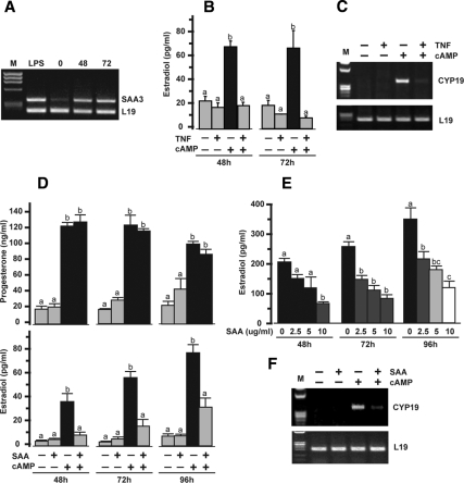
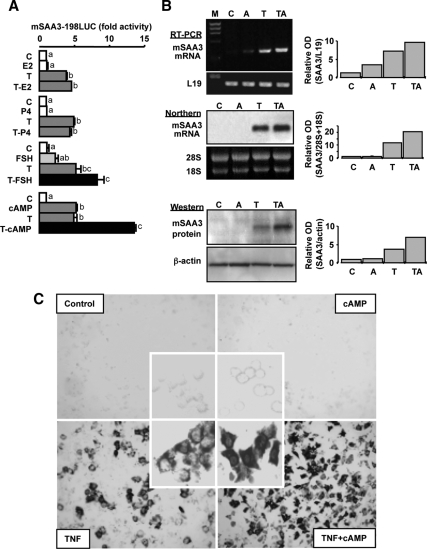
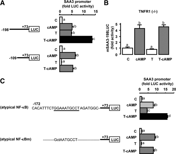
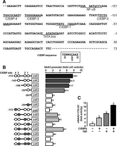
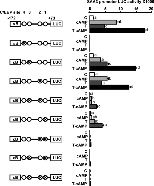
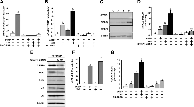
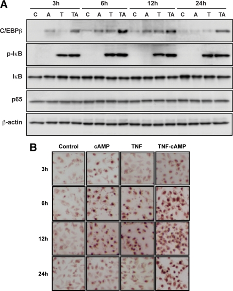
TNFalpha is an inflammatory-related cytokine that has inhibitory effects on gonadotropin- and cAMP-stimulated steroidogenesis and folliculogenesis. Because ovulation is an inflammatory reaction and TNF specifically induces serum amyloid A3 (SAA3) in mouse granulosa cells, the effect of cAMP on TNF-induced SAA3 promoter activity, mRNA and protein was investigated. Granulosa cells from immature mice were cultured with TNF and/or cAMP. TNF increased SAA3 promoter activity, mRNA, and protein, which were further increased by cAMP. cAMP alone increased SAA3 promoter activity, but SAA3 mRNA and protein remained undetectable. Thus, there appeared to be different mechanisms by which TNF and cAMP regulated SAA3 expression. SAA3 promoters lacking a nuclear factor (NF)-kappaB-like site or containing its mutant were not responsive to TNF but were responsive to cAMP. Among four CCAAT-enhancing binding protein (C/EBP) sites in the SAA3 promoter, the C/EBP site nearest the NF-kappaB-like site was required for TNF-induced SAA3. The C/EBP site at -75/-67 was necessary for responsiveness to cAMP. Dominant-negative C/EBP and cAMP response element-binding protein or short interfering RNA of C/EBPbeta blocked TNF- or cAMP-induced SAA3 promoter activity. The combination of TNF and cAMP increased C/EBPbeta protein above that induced by TNF or cAMP alone. Thus, cAMP in combination with TNF specifically induced C/EBPbeta protein, leading to enhanced SAA3 expression but requiring NF-kappaB in mouse granulose cells. In addition, like TNF, SAA inhibited cAMP-induced estradiol accumulation and CYP19 levels. These data indicate SAA may play a role in events occurring during the ovulation process.


Figures

Figure 1
Figure 1
Effects of TNF and SAA on mouse granulosa cell progesterone and estradiol production and CYP19 mRNA expression. A, Effects of eCG and hCG administration on mouse ovarian SAA3 mRNA. Ovaries were collected 0, 48, and 72 h after eCG (2.5 IU); hCG was administered 52 h after eCG. Ovaries were collected from LPS-treated mice and serve as a positive control. L19 was used as a loading control for RT-PCR. M, Molecular marker. B, Effects of TNF (10 ng/ml) in the presence and absence of cAMP (500 μm) on estradiol production after 48 and 72 h. C, Effects of TNF (10 ng/ml) in the presence and absence of cAMP (500 μm) on granulosal CYP19 expression after 72 h. L19 was used as a loading control for RT-PCR. M, Molecular marker. D, Effects of SAA (10 μg/ml) in the presence and absence of cAMP (500 μm) on progesterone and estradiol production after 48, 72, and 96 h. E, Dose-dependent effects of SAA (0–10 μg/ml) on cAMP (500 μm)-stimulated estradiol production after 48, 72, and 96 h. F, Effects of SAA (10 μg/ml) in the presence and absence of cAMP (500 μm) on CYP19 expression after 96 h. L19 was used as a loading control for RT-PCR. M, Molecular marker. Bars with different colors and letters are significantly different (P ≤ 0.05) by ANOVA and Tukey’s test.
Figure 2
Figure 2
Effects of hormones and cAMP on TNF-induced SAA3. Panel A, Effects of estradiol, progesterone, FSH, and cAMP on TNF-induced SAA3 promoter (mSAA3-198LUC) activity in mouse granulosa cells. Cells were transfected for 3.5 h with SAA3 promoter luciferase reporter and then incubated with vehicle or TNF (10 ng/ml) in the absence or presence of steroids [estradiol (E2), 1 μm; progesterone (P4), 1 μm], FSH (10 ng/ml), or cAMP (500 μm) for 16 h. Luciferase activity was normalized using total protein concentrations and expressed as a fold increase compared with the control. Bars with different colors and letters are significantly different (P ≤ 0.05) by ANOVA and Tukey’s test. C, Control; T, TNF; LUC, luciferase. Panel B, Effect of cAMP on TNF-induced SAA3 mRNA and protein in mouse granulosa cells. After isolating total RNAs and proteins, RT-PCR, Northern and Western blots were performed using specific primers, cDNA probe, and antibody for SAA3, respectively. Loading controls, L19, 28S, and 18S, and β-actin were used for RT-PCR and Northern and Western blot, respectively. These figures represent data from duplicate experiments. M, Molecular marker; C, control; A, cAMP; T, TNF; TA, TNF+cAMP. Panel C, Effect of cAMP on TNF-induced SAA3 mRNA in cultured mouse granulosa cells determined by in situ hybridization.
Figure 3
Figure 3
Differential mechanisms between cAMP and TNF in regulating SAA3 promoter activity. A, Effects of cAMP on the luciferase activity of TNF-induced SAA3 promoters (mSAA3-198LUC and mSAA3-109LUC) in mouse granulosa cells. B, Effects of cAMP and TNF on SAA3 promoter (mSAA3-198LUC) activity in granulosa cells cultured from p55 TNF receptor type 1 (TNFR1) knockout mice (−/−). C, Effects of cAMP and TNF on the activity of SAA3 promoters containing an atypical NF-κB binding site and its mutant in mouse granulosa cells. Cells were transfected for 3.5 h with various mSAA3 promoters and then were incubated with or without TNF (10 ng/ml) in the absence or presence of cAMP (500 μm) for 16 h. Luciferase activity was normalized using total protein concentrations and expressed as a fold change in comparison with the control. Bars with different colors and letters are significantly different (P ≤ 0.05) by ANOVA and Tukey’s test. Atypical NF-κB sequence is underlined and its mutant (atypical NF-κBm) is indicated by lowe-case letters. C, Control; T, TNF; LUC, luciferase.
Figure 4
Figure 4
Involvement of C/EBP sites in cAMP-regulated SAA3 promoter activity. A, Nucleotide sequence of SAA3 promoter (−198/+73). The SAA3 promoter contained one atypical NF-κB and four C/EBP sites. Lowercase letters indicate base pairs different from consensus sites. B, Effects of cAMP on the activity of SAA3 promoters containing serially deleted and mutated C/EBP sites in mouse granulosa cells. Data for each promoter is fold change with cAMP treatment compared with control; statistical significance (P ≤ 0.05) by Student’s t test is indicated by a colored bar. Comparison of camp-stimulated activity between promoters was by ANOVA and Tukey’s test, and bars with different colors and numbers are statistically different (P ≤ 0.05). C, Effects of C/EBPβ on TNF-induced SAA3 promoter (mSAA3-172LUC) activity in mouse granulosa cells. Cells were transfected for 3.5 h with mSAA3 promoter with or without a C/EBPβ expression vector (100 ng/ml) and then incubated with or without cAMP (500 μm) for 16 h. Luciferase activity was normalized to total protein concentration and expressed as a fold change compared with the control. Bars with different colors and letters are significantly different (P ≤ 0.05) by ANOVA and Tukey’s test. Cross and open circles indicate mutated and normal C/EBP sites, respectively. LUC, Luciferase.
Figure 5
Figure 5
Identification of C/EBP sites regulating SAA3 promoter activity. Interaction of cAMP and TNF on the activity of mSAA3-172LUC containing mutated C/EBP sites in mouse granulosa cells is shown. Cross and open circles indicate mutated and intact C/EBP sites, respectively. Cells were transfected for 3.5 h with mSAA3 promoters and then incubated with vehicle or TNF (10 ng/ml) in the absence or presence of cAMP (500 μm) for 16 h. Luciferase activity is reported and was normalized to total protein concentrations. Bars with different colors and letters are significantly different (P ≤ 0.05) by ANOVA and Tukey’s Test. κB, NF-κB site; C, control; T, TNF; LUC, luciferase.
Figure 6
Figure 6
Effects of dominant-negative C/EBP, C/EBPβ siRNA, and dominant-negative CREB on cAMP-, TNF-, and p65-induced SAA3 promoter activity. Panel A, Effect of dominant-negative C/EBP on TNF-induced SAA3 promoter activity in mouse granulosa cells. DN, Dominant-negative. Panel B, Effect of dominant-negative C/EBP on interaction between cAMP and p65 in increasing SAA3 promoter activity. Cells were transfected for 3.5 h with mSAA3-172LUC alone or with dominant-negative C/EBP vector (100 ng/ml) and p65 expression vector (10 ng/ml) and then incubated with TNF (10 ng/ml) alone and/or with cAMP (500 μm) for 16 h. The luciferase activity was normalized using total protein concentrations and expressed as a fold change in comparison with the control. DN, Dominant negative; LUC, luciferase. Panel C, Effects of TNF and cAMP on C/EBP isoforms in mouse granulosa cells. Cells were cultured with TNF (10 ng/ml) and cAMP (500 μm) alone and in combination for 24 h. Each lane was loaded with 20 μg of total protein using whole-cell lysates. β-Actin was used as a loading control. C, Control; A, cAMP; T, TNF; TA, TNF+cAMP. Panel D, Inhibitory effect of C/EBPβ siRNA on cAMP and TNF in enhancing SAA3 promoter activity. Cells were transfected for 24 h with C/EBPβ siRNA (10 nm) and then further transfected for 3.5 h with mSAA3-172LUC. Transfected cells were incubated with TNF (10 ng/ml) alone, cAMP (500 μm) alone, and in combination for 16 h. Panel E, Effect of C/EBPβ siRNA on C/EBPβ, SAA3, phospho-IκB, IκB, and p65 protein expression after cAMP and TNF treatments in mouse granulosa cells. Cells were transfected for 24 h with C/EBPβ siRNA (10 nm) and then incubated with TNF (10 ng/ml) and cAMP (500 μm) in combination for 24 h. Panel F, Effect of cAMP and TNF on pCRE-luc promoter activity in granulosa cells. Cells were transfected for 3.5 h with pCRE-luc and then incubated with TNF (10 ng/ml) alone, cAMP (500 μm) alone, and in combination for 16 h. Luciferase activity was normalized using total protein concentrations and expressed as a fold change in comparison with the control. Panel G, Effect of dominant-negative CREB on interaction between cAMP and TNF in increasing SAA3 promoter activity. Cells were transfected for 3.5 h with mSAA3–172LUC alone or with dominant-negative CREB vector (100 ng/ml) and then incubated with TNF (10 ng/ml) alone and/or with cAMP (500 μm) for 16 h. Luciferase activity was normalized using total protein concentrations and expressed as a fold change in comparison with the control. Different color bars indicate significant differences (P ≤ 0.05) among groups. DN, Dominant-negative; LUC, luciferase.
Figure 7
Figure 7
Time course of C/EBPβ, phospho-IκB (p-IκB), IκB, and p65 proteins in response to cAMP and TNF. Panel A, Effects of cAMP and TNF on the time course of C/EBPβ, p-IκB, IκB, and p65 proteins in mouse granulosa cells. Cells were incubated with TNF (10 ng/ml) alone, cAMP (500 μm) alone, and in combination for 3, 6, 12, and 24 h. Each gel lane was loaded with 20 μg of total protein using whole-cell lysates. β-Actin was used as a loading control. Panel B, Nuclear localization of C/EBPβ protein by immunocytochemistry after cAMP and/or TNF in mouse granulosa cells. The experiments were repeated twice and a representative figure is shown. C, Control; A, cAMP; T, TNF; TA, TNF+cAMP.

Similar articles

Cited by

References

    1. Bagavandoss P, Kunkel SL, Wiggins RC, Keyes PL 1988 Tumor necrosis factor-α (TNF-α) production and localization of macrophages and T lymphocytes in the rabbit corpus luteum. Endocrinology 122:1185–1187 - PubMed
    1. Barak V, Mordel N, Holzer H, Zajicek G, Treves AJ, Laufer N 1992 The correlation of interleukin 1 and tumor necrosis factor to oestradiol, progesterone and testosterone levels in periovulatory follicular fluid of in vitro fertilization patients. Hum Reprod 7:462–464 - PubMed
    1. Chen HL, Marcinkiewicz JL, Sancho-Tello M, Hunt JS, Terranova PF 1993 Tumor necrosis factor α gene expression in mouse oocytes and follicular cells. Biol Reprod 48:707–714 - PubMed
    1. Hehnke-Vagnoni KE, Clark CL, Taylor MJ, Ford SP 1995 Presence and localization of tumor necrosis factor-α in the corpus luteum of nonpregnant and pregnant pigs. Biol Reprod 53:1339–1344 - PubMed
    1. Ji I, Slaughter RG, Ellis JA, Ji TH, Murdoch WJ 1991 Analyses of ovine corpora lutea for tumor necrosis factor mRNA and bioactivity during prostaglandin-induced luteolysis. Mol Cell Endocrinol 81:77–80 - PubMed

Publication types

MeSH terms

Substances