. 2010 Jul 1;21(13):2217-25.
doi: 10.1091/mbc.e10-01-0081. Epub 2010 May 5.

The polarized distribution of Na+,K+-ATPase: role of the interaction between {beta} subunits

Affiliations

The polarized distribution of Na+,K+-ATPase: role of the interaction between {beta} subunits

Teresita Padilla-Benavides et al. Mol Biol Cell. .

Abstract

The very existence of higher metazoans depends on the vectorial transport of substances across epithelia. A crucial element of this transport is the membrane enzyme Na(+),K(+)-ATPase. Not only is this enzyme distributed in a polarized manner in a restricted domain of the plasma membrane but also it creates the ionic gradients that drive the net movement of glucose, amino acids, and ions across the entire epithelium. In a previous work, we have shown that Na(+),K(+)-ATPase polarity depends on interactions between the beta subunits of Na(+),K(+)-ATPases located on neighboring cells and that these interactions anchor the entire enzyme at the borders of the intercellular space. In the present study, we used fluorescence resonance energy transfer and coprecipitation methods to demonstrate that these beta subunits have sufficient proximity and affinity to permit a direct interaction, without requiring any additional extracellular molecules to span the distance.


Figures

Figure 1.
Figure 1.
MDCK cells express β1 subunits at homotypic but not at heterotypic contacts. Localization of the dog β1 subunit (A–C, green) of Na+,K+-ATPase was studied by immunofluorescence assay in MDCK cells derived from dog kidney. (A) Monolayer of pure MDCK cells showing that the β1 subunit is only expressed at the plasma membranes in the lateral domain where cells contact each other. (B) MDCK cells cocultured with NRK cells (derived from normal rat kidney) that were labeled beforehand with CMTMR (red). In the mixed monolayer, the β1 subunit is only expressed at homotypic borders (MDCK/MDCK) but not at heterotypic ones (MDCK/NRK). (C) Monolayer of mixed MDCK/NRK cells transfected with dog β1 subunit shows that the β subunit (green) is concentrated at both homotypic MDCK/MDCK contacts and heterotypic MDCK/NRK contacts. Bars, 20 μm.
Figure 2.
Figure 2.
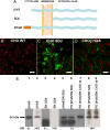
β Subunits interact in vitro. (A) Scheme of the wild-type and recombinant β subunits used in this study. Shown are the wild-type β subunit (βWT, top), the soluble β subunit consisting only of the extracellular domain (SDβ, middle), and the β subunit tagged with a hexahistidine repeat (βHis6, bottom). (B–D) Immunofluorescence images of the dog β1 subunit expressed in CHO cells. CHO WT cells (B), CHO cells expressing SDβ (C), and CHO cells expressing βHis6 in their plasma membranes (D) are depicted. Nuclei were stained with propidium iodide (red). Bars, 40, 30, and 20 μm, respectively. (E) Pull-down assay analysis. Western blots using an antibody against the dog β1 subunit and a His probe reveal, in both cases, the recombinant βHis6 protein (lane 1 and 2), whereas only the dog β1 antibody recognizes the soluble extracellular domain of dog β1 (SDβ, lanes 3 and 4). A mixture of purified SDβ with βHis6 produces the mixed pattern shown in lane 5. The proteins eluted from a pull-down (PD) assay by using βHis6 as bait and soluble SDβ as prey (lane 6) show the same pattern of bands. Only the band corresponding to the bait protein (βHis6) is observed when the pull-down is performed with CHO WT cell supernatant (lane 7). This is confirmed when both pull-downs are blotted to detect the His6 repeat (lanes 8 and 9).
Figure 3.
Figure 3.
Cellular expression of molecular tools. (A) Scheme of the β subunits fused to donor or acceptor fluorophores. Shown are the rat β1 subunit fused to CFP in the extracellular domain (Rβ CFP, top) rat β1 subunit fused to YFP in the extracellular domain (Rβ YFP). (B–D) Western blot assays to detect the expression of both constructs in transfected MDCK cells (MRβ CFP or MRβ YFP). (B) Detection of β1 subunits with a specific antibody against the dog subunit. No band is detected in rat NRK-E52 cells. (C) Detection of the rat β1 subunit with a specific antibody for this species. No band corresponding to the rat β1 subunit is observed in WT MDCK cells. (D) Immunodetection using an anti-GFP antibody. A 95-kDa band corresponding to the fluorescent constructs (MRβ CFP or MRβ YFP) is visible. This band coincides with the band recognized by the rat-specific antibody and does not appear in MDCK WT cells. (E–H) Confocal images of WT MDCK cells and MDCK cells transfected with Rβ CFP or YFP. (E) IF of the endogenous dog β1 and rat β1 (F) subunits in WT MDCK cells. (G) Fluorescence imaging of Rβ CFP obtained by exciting at 390–425 nm and recording its recovery at 475–525 nm. (H) Fluorescence imaging of the Rβ YFP obtained by exciting at 475–515 nm and recording its emission at 540–575 nm. Bar, 20 μm.
Figure 4.
Figure 4.
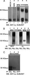
Interaction between β subunits of neighboring cells. (A) Immunodetection of β1 subunits with the pan-species β1 subunit antibody. Characteristic bands of MDCK WT (lane 1), NRK WT (lane 2), and cocultures of NRK WT and MRβ YFP cells (lane 3) blotted with the pan-species β1 subunit antibody, dog (Dβ1) and rat subunits (Rβ1). The low-molecular-weight endogenous β1 and the high-molecular-weight β1-YFP are both recognized by the same antibody (lane 4). Immunoprecipitated cocultures of NRK WT and MRβ YFP cells by using an antibody against GFP reveal a band corresponding to the recombinant Rβ YFP (95-kDa band; lane 5) and a coimmunoprecipitated lighter band corresponding to the native β1 subunit (55-kDa band in lane 5). (B) Immunoprecipitation with an anti-GFP antibody shows no bands when blotted with an antibody against Dβ1 (lane 1). If the blot is instead performed with the pan-species β1 subunit antibody, both the endogenous and recombinant proteins are revealed (lane 2). The same pattern is seen if the immunoprecipitations are performed with the pan-species β1 antibody (lanes 3 and 4). Immunoprecipitation with the β1 antibody of MDCK cells expressing recombinant rat β1 (MRβ YFP) shows the endogenous β1 (lane 5) when blotted with the Dβ1 antibody and the transfected one when detected with the Rβ1 antibody (lane 6). (C) As negative controls, we immunoprecipitated cocultures of MDCK WT with NRK cells and probed them with Dβ1, Rβ1, and GFP antibodies. As expected, the endogenous dog (lane 1) and rat (lane 2) subunits were detected, whereas YFP-β1 was not (lane 3).
Figure 5.
Figure 5.
Interaction between β1 subunits observed by FRET after the in vivo acceptor photobleaching assay. (A) Fluorescence image captured before acceptor photobleaching in cocultures of MRβ CFP (cyan) and MRβ YFP (yellow) living cells. The optical merge shows the colocalization of both fluorescent proteins in cell-cell contact areas (green). (B) Fluorescence of the same optical section after photobleaching of the YFP in a membrane section that possessed both CFP and YFP expression. ROI 1 was the region used for quantification of %E. Bleaching of YFP in this region increased the fluorescence of CFP. ROI 2 was the region used as an internal negative control, in which the change of CFP fluorescence was also measured. Bar, 20 μm. (C) Quantification of %E. The percentage of energy transference obtained in 10 experiments was averaged. Bars represent SEM and asterisks indicate p < 0.001 with respect to negative control. ROI 3 is from MDCK cells expressing a CFP-YFP tandem construct (positive control) and ROI 1 and 2 are the same as in B. Data are summarized in Supplemental Table 1.
Figure 6.
Figure 6.
Polarized expression of epithelial Na+,K+-ATPase. (A) Seminal model of Koefoed-Johnsen and Ussing (1958), in which the Na+,K+-ATPase (reinforced in magenta) was assumed to occupy the basal side of the cell, which constitutes the inner-facing barrier. (B) From this position, the pump transports Na+ toward the interstitial side of the cell, producing a net decrease in the cytoplasmic concentration of this ion and setting up an electrochemical force that drives counter- and cotransporters of sugars, amino acids, and ions and possible the existence of metazoans. (C) Confocal transverse section of a monolayer of MDCK cells. The nuclei are stained with propidium iodide (red) and the β subunits of Na+,K+-ATPase are stained with a specific antibody (green), showing that this subunit is localized to the lateral surfaces of cells, but not to the apical (left) or basal sides. (D) Na+,K+-ATPase molecules of two neighboring epithelial cells with interacting β subunits (green), as shown previously (Shoshani et al., 2005) and in the present work. (E) Na+,K+-ATPase molecules anchored to the lateral membranes. Because of the tight junction, Na+ ions pumped into the intercellular space can only diffuse inwards, generating vectorial transport across the epithelium.

Similar articles

Cited by

References

    1. Arrate M. P., Rodriguez J. M., Tran T. M., Brock T. A., Cunningham S. A. Cloning of human junctional adhesion molecule 3 (JAM3) and its identification as the JAM2 counter-receptor. J. Biol. Chem. 2001;276:45826–45832. - PubMed
    1. Bab-Dinitz E., Albeck S., Peleg Y., Brumfeld J., Gottschalk K. E., Karlish S. A C-terminal lobe of the β subunit of Na, K-ATPase and H, K-ATPase resembles cell adhesion molecules. Biochemistry. 2009;48:8684–8691. - PubMed
    1. Belusa R., Wang Z., Matsubara T., Sahingren B., Dulubova I., Nair A. C., Ruoslathi E., Greengard P., Aperia A. “Mutation of the protein kinase C phosphorylation site on rat α1 Na+, K+-ATPase alters regulation of intracellular Na+ and pH and influences Cell shape and adhesiveness”. J. Biol. Chem. 1997;272:20179–20184. - PubMed
    1. Blasig I. E., Winkler L., Lassowski B., Mueller S. L., Zuleger N., Krause E., Krause G., Gast K., Kolbe M., Piontek J. On the self-association potential of transmembrane tight junction proteins. Cell. Mol. Life. Sci. 2006;63:505–514. - PMC - PubMed
    1. Caplan M. J., Anderson H. C., Palade G. E., Jamieson J. D. Intracellular sorting and polarized cell surface delivery of (Na+, K+)ATPase, an endogenous component of MDCK cell basolateral plasma membranes. Cell. 1986;46:623–631. - PubMed

Publication types

MeSH terms

LinkOut - more resources