. 2010 Jul 1;21(13):2202-16.
doi: 10.1091/mbc.e09-11-0962. Epub 2010 May 5.

Fusel alcohols regulate translation initiation by inhibiting eIF2B to reduce ternary complex in a mechanism that may involve altering the integrity and dynamics of the eIF2B body

Affiliations

Fusel alcohols regulate translation initiation by inhibiting eIF2B to reduce ternary complex in a mechanism that may involve altering the integrity and dynamics of the eIF2B body

Eleanor J Taylor et al. Mol Biol Cell. .

Abstract

Recycling of eIF2-GDP to the GTP-bound form constitutes a core essential, regulated step in eukaryotic translation. This reaction is mediated by eIF2B, a heteropentameric factor with important links to human disease. eIF2 in the GTP-bound form binds to methionyl initiator tRNA to form a ternary complex, and the levels of this ternary complex can be a critical determinant of the rate of protein synthesis. Here we show that eIF2B serves as the target for translation inhibition by various fusel alcohols in yeast. Fusel alcohols are endpoint metabolites from amino acid catabolism, which signal nitrogen scarcity. We show that the inhibition of eIF2B leads to reduced ternary complex levels and that different eIF2B subunit mutants alter fusel alcohol sensitivity. A DNA tiling array strategy was developed that overcame difficulties in the identification of these mutants where the phenotypic distinctions were too subtle for classical complementation cloning. Fusel alcohols also lead to eIF2alpha dephosphorylation in a Sit4p-dependent manner. In yeast, eIF2B occupies a large cytoplasmic body where guanine nucleotide exchange on eIF2 can occur and be regulated. Fusel alcohols impact on both the movement and dynamics of this 2B body. Overall, these results confirm that the guanine nucleotide exchange factor, eIF2B, is targeted by fusel alcohols. Moreover, they highlight a potential connection between the movement or integrity of the 2B body and eIF2B regulation.


Figures

Figure 1.
Figure 1.
Fusel alcohols differentially inhibit translation initiation in butanol-sensitive (BUTS) and -resistant (BUTR) strains. (A) Polysome traces from yMK23 (BUTR) and yMK36 (BUTS) strains grown in YPD and incubated with added 1-butanol at the concentrations indicated for 10 min at 30°C. Polysomes were analyzed as described in Materials and Methods. The 40S (small ribosomal subunit), 60S (large ribosomal subunit), 80S (monosome), and polysome peaks are labeled. (B) Polysome traces from the yMK23 (BUTR) and yMK36 (BUTS) strains as in A, except the yeast were incubated with the indicated concentrations of tert-amyl alcohol (2-methyl 2-butanol), isobutyl alcohol (2-methyl 1-propanol), and isoamyl alcohol (3-methyl 1-butanol) before polysome analysis.
Figure 2.
Figure 2.
Parallel mutations within the left-handed parallel β-helix (LbH) domains of eIF2Bε and γ lead to opposite phenotypes after exposure to butanol. (A) Polysome traces from yMK658 (GCD6) or yMK644 (gcd6-1) strains, grown in YPD and treated with butanol at the indicated concentrations for 10 min at 30°C. (B) Polysome traces from yMK974 (GCD1) or yMK976 (GCD1-C483W) strains, grown in selective media and treated with butanol at the indicated concentrations for 10 min at 30°C. Polysome analysis was as described in Materials and Methods.
Figure 3.
Figure 3.
DNA tiling arrays highlight single nucleotide polymorphisms (SNPs) in the α subunit of eIF2B, which may explain the resistance to butanol. (A) Gradient plate and polysome analysis on yMK54 (BUT1-1), yMK16 (BUT1-2), and yMK36 (progenitor) showing the resistant profiles of the mutants relative to the progenitor BUTS strain. The gradient plates contain a gradient of butanol up to a maximum of 3% vol/vol, and equal numbers of the yeast cells are spotted at every position. (B) The five SNPs identified as present in yMK16 (BUT1-2) strain but not in yMK36 (progenitor) are shown along with the genomic position, i.e., chromosome number and coordinate. These include a SNP in the α subunit of eIF2B (GCN3). (C) A plot showing the SNP prediction signal across the GCN3 gene locus obtained from the tiling microarray analysis. A stringent cutoff value of 10 was used, and a prominent potential SNP was identified for the yMK16 (BUT1-2) strain (red) but not for the yMK36 (progenitor) strain (green). Sequencing analysis of multiple independently PCR-amplified DNA samples across the GCN3 locus identified a single amino acid change of arginine to lysine at position 148 for the yMK16 (BUT1-2) strain relative to the progenitor.
Figure 4.
Figure 4.
Specific point mutations but not deletions of the eIF2Bα gene are resistant to butanol treatment. (A) Polysome traces from yMK36 (GCD1-S180) or yMK1262 (GCD1-S180 gcn3Δ) strains, grown in selective media and incubated either in amino acid–free media for 15 min or in media containing butanol at the indicated concentrations for 10 min at 30°C. (B) Polysome traces from gcn3Δ strains bearing various plasmids, yMK1442 with p[GCN3 URA3 CEN], yMK1441 with p[GCN3-R148K URA3 CEN], and yMK1451 with p[GCN3-T41K URA3 CEN]. Strains were grown in selective media and incubated in fresh media or media containing butanol at the indicated concentrations for 10 min at 30°C. Polysome analysis was carried out as described in Materials and Methods. (C) Gradient plate analysis on the above strains showing the resistant profiles of the GCN3 mutant strains relative to the wild type. The gradient plates contain a gradient of butanol up to a maximum of 3% vol/vol, and equal levels of yeast are spotted at every position.
Figure 5.
Figure 5.
Butanol causes a Sit4p-dependent dephosphorylation of eIF2α. (A) Western blots from the yMK23 [GCD1-P180 (BUTR); top panels] and yMK36 [GCD1-S180 (BUTS); bottom panels] strains after growth in SCD. Cells were either untreated (UT) or treated for 15 min in the indicated concentrations of alcohol. Cells were also pelleted and resuspended in complete media (SCD) or media lacking amino acids (SCD-AAs) for 15 min. Protein extracts were blotted and probed with antibodies to eIF2α and phospho-specific antibodies to phosphoserine 51 on eIF2α. (B) As in A, except the strains were pretreated in amino acid starvation media for 15 min to induce eIF2α phosphorylation before 1% (vol/vol) butanol addition. UT, untreated cells; 1, the pretreatment alone, 2 through 5, 5, 10, 15, and 30 min of butanol addition under continuing amino acid starvation conditions; 6, a control of 30-min amino acid starvation alone after the pretreatment. (C) Protein extracts were made from yMK472 (BY4741 wild type) and a range of phosphatase subunit mutants (yMK1235-49) to screen for mutants that were defective in the butanol-induced dephosphorylation of eIF2α. After growth on SCD media, extracts were made from untreated cells (UT), cells starved for amino acids for 15 min (−AAs), and cells treated with 2% (vol/vol) butanol after the amino acid starvation (−AAs +But). (D) Polysome traces from yMK472 (BY4741) and yMK1246 (BY4741 sit4Δ) strains, grown in rich media and incubated in media containing butanol at the indicated concentrations for 10 min at 30°C.
Figure 6.
Figure 6.
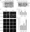
Butanol reduces ternary complex levels and modestly reduces the transit of eIF2 through the 2B body. (A) Myc-immunoprecipitation experiments using extracts prepared from the yMK1125 [bearing GCD1-P180-MYC (BUTR)] and yMK1126 [bearing GCD1-S180-MYC (BUTS)] strains grown in rich media and then either incubated in media (−) or media with 1% (vol/vol) butanol (+) for 15 min. Samples were separated into input and pellet fractions, as indicated, and were used for Western blots using antibodies against eIF2Bα to ε. (B) Flag-immunoprecipitation experiments using extracts prepared from strains yMK1613 [GCD1-P180 (BUTR)] and yMK1615 [GCD1-S180 (BUTS)] (both bearing a eIF2β-Flag–expressing plasmid) grown in rich media and then either incubated in media (−) or media with 1% (vol/vol) butanol (+) for 15 min. Samples were separated into input and pellet fractions, as indicated, and 20% was used for Western blots using antibodies against Flag, eIF2α to eIF2γ. The remaining 80% was used to prepare RNA that was used to perform Northern blots using an oligonucleotide probe to tRNAiMet. (C) A panel of live-cell images from the BUTR (GCD1-P180) strains: yMK883 (eIF2α-GFP), yMK1211 (eIF2γ-GFP), yMK880 (eIF2Bγ-GFP), yMK882 (eIF2Bε-GFP), yMK1347 (eIF2Bβ-GFP), yMK1356 (eIF2Bα-GFP) and the BUTS (GCD1-S180) strains: yMK914 (eIF2α-GFP), yMK1212 (eIF2γ-GFP), yMK876 (eIF2Bγ-GFP), yMK878 (eIF2Bε-GFP), yMK1363 (eIF2Bβ-GFP), and yMK1355 (eIF2Bα-GFP) either treated with 1% (vol/vol) butanol for 15 min (+) or untreated (−). (D) Bar chart depicting the mean percentage eIF2 in the 2B body for the strains yMK883 (SUI2-GFP GCD1-P180 BUTR) and yMK914 (SUI2-GFP GCD1-S180 BUTS) after 15-min treatments in complete media (−), media with l% (vol/vol) butanol (+but), or media lacking amino acids (−AAs). eIF2 levels in the 2B body were measured using confocal microscopy and densitometry. Fifteen image-merged z-stacks from at least 20 single yeast cells were quantified using ImageJ to obtain the plotted values. Error bars, ±1 SEM; *p < 0.01 from an analysis of variance (ANOVA). (E) Bar chart showing half times of FRAP derived from experiments performed on eIF2α-GFP–bearing strains yMK883 (SUI2-GFP GCD1-P180 BUTR) and yMK914 (SUI2-GFP GCD1-S180 BUTS). Strains were grown in SCD media then incubated for 15 min in either complete media (−), media with l% (vol/vol) butanol (+but), or media lacking amino acids (−AAs) before single-cell FRAP studies. Error bars, ±1 SEM (where n is at least 18). *p < 0.05 from analysis of variance (ANOVA).
Figure 7.
Figure 7.
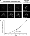
The 2B body rapidly moves throughout the cell. (A) Images from time-lapse microscopy studies using the yMK880 (eIF2Bγ-GFP) and yMK883 (eIF2α-GFP) strains in the top and bottom rows, respectively. Each row contains three stills from a series of 25 images over a period of 2 min as well as a merged image of all 25 stills, which serves to depict the total extent of 2B body movement. (B) A plot of the mean square displacement (MSD) as a function of time interval (Δt) from >25 single particle–tracking experiments using the yMK880 (eIF2Bγ-GFP) strain. For short time intervals the 2B body does not become displaced linearly, suggesting that there is some form of tethering in operation. At progressively longer time intervals the MSD approaches a linear relationship, which is indicative of movement by diffusion.
Figure 8.
Figure 8.
Butanol inhibits 2B body movement and butanol-resistant GCN3 mutants lack the 2B body. (A) Images from time-lapse microscopy studies using the eIF2Bγ-GFP bearing strains yMK876 [GCD1-S180-GFP (BUTS)] and yMK880 [GCD1-P180-GFP (BUTR)] strains, top three rows and bottom three rows, respectively. The strains were incubated in media (−) or media with 1% (vol/vol) or 2% (vol/vol) butanol for 10 min as indicated. Each row contains three stills from a series of 25 images over a period of 2 min, as well as a merged image of all 25 stills, which serves to depict the total extent of 2B body movement. (B) Bar chart depicting the mean distance moved in μ over a 2-min period from at least 40 time-lapse experiments (as in A) using eIF2α-GFP to mark the 2B body in the strains yMK914 [SUI2-GFP GCD1-S180 (BUTS)] and yMK883 [SUI2-GFP GCD1-P180 (BUTR)] Error bars, ±1 SEM; *p < 0.01 from an analysis of variance (ANOVA). (C) As in B, except eIF2Bγ is GFP-tagged to follow the 2B body in the strains yMK876 [GCD1-S180-GFP (BUTS)] and yMK880 [GCD1-P180-GFP (BUTR)]. (D) Images showing the absence of the eIF2B body in the yMK1567 [BUT1-1 (GCN3-R148K)] and yMK1595 [(BUT1-2 (GCN3-T41A)] strains relative to wild-type controls yMK1596 (GCD1-P180 BUTR) and yMK1597 (GCD1-S180 BUTS) using eIF2Bε-GFP (GCD6-GFP) to mark the eIF2B body.

Similar articles

Cited by

References

    1. Aravind L., Koonin E. V. Eukaryote-specific domains in translation initiation factors: implications for translation regulation and evolution of the translation system. Genome Res. 2000;10:1172–1184. - PMC - PubMed
    1. Ashe M. P., De Long S. K., Sachs A. B. Glucose depletion rapidly inhibits translation initiation in yeast. Mol. Biol. Cell. 2000;11:833–848. - PMC - PubMed
    1. Ashe M. P., Slaven J. W., De Long S. K., Ibrahimo S., Sachs A. B. A novel eIF2B-dependent mechanism of translational control in yeast as a response to fusel alcohols. EMBO J. 2001;20:6464–6474. - PMC - PubMed
    1. Burns N., Grimwade B., Rossmacdonald P. B., Choi E. Y., Finberg K., Roeder G. S., Snyder M. Large-scale analysis of gene expression, protein localization, and gene disruption Saccharomyces cerevisiae. Gene Dev. 1994;8:1087–1105. - PubMed
    1. Bushman J. L., Asuru A. I., Matts R. L., Hinnebusch A. G. Evidence that GCD6 and GCD7, translational regulators of GCN4, are subunits of the guanine nucleotide exchange factor for eIF-2 in Saccharomyces cerevisiae. Mol. Cell. Biol. 1993a;13:1920–1932. - PMC - PubMed

Publication types

MeSH terms

LinkOut - more resources