. 2010 Jul 1;21(13):2270-84.
doi: 10.1091/mbc.e09-04-0345. Epub 2010 May 5.

Exit from the Golgi is required for the expansion of the autophagosomal phagophore in yeast Saccharomyces cerevisiae

Affiliations

Exit from the Golgi is required for the expansion of the autophagosomal phagophore in yeast Saccharomyces cerevisiae

Aniek van der Vaart et al. Mol Biol Cell. .

Abstract

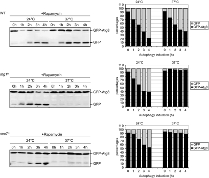
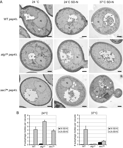
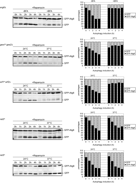
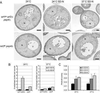
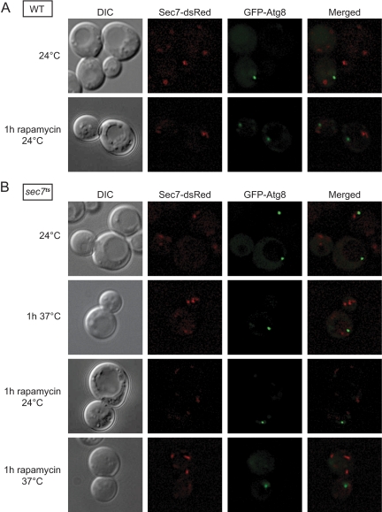
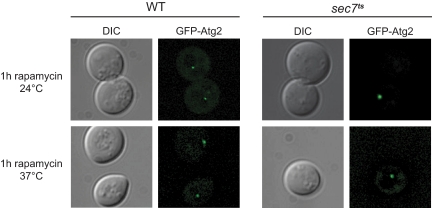
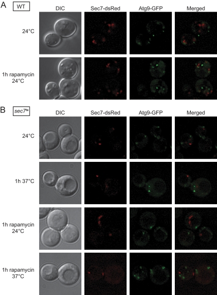
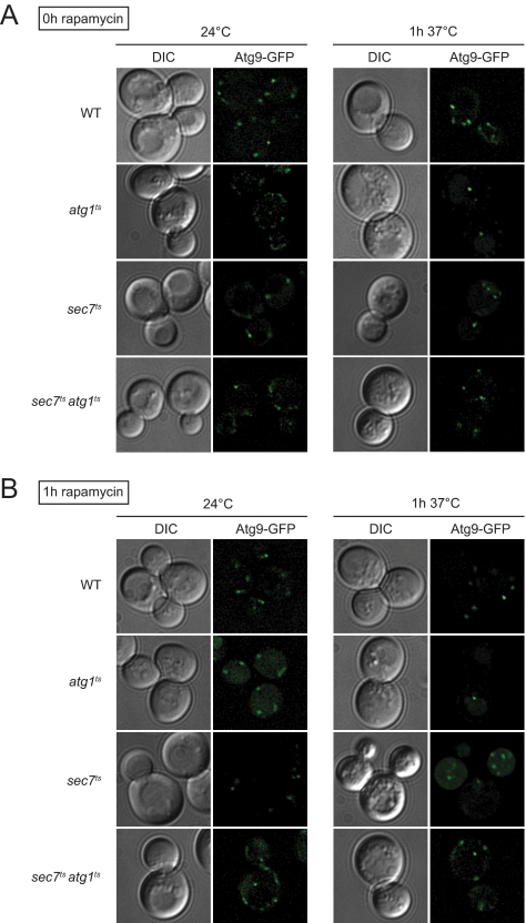
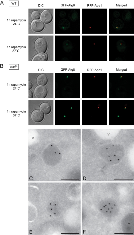
The delivery of proteins and organelles to the vacuole by autophagy involves membrane rearrangements that result in the formation of large vesicles called autophagosomes. The mechanism underlying autophagosome biogenesis and the origin of the membranes composing these vesicles remains largely unclear. We have investigated the role of the Golgi complex in autophagy and have determined that in yeast, activation of ADP-ribosylation factor (Arf)1 and Arf2 GTPases by Sec7, Gea1, and Gea2 is essential for this catabolic process. The two main events catalyzed by these components, the biogenesis of COPI- and clathrin-coated vesicles, do not play a critical role in autophagy. Analysis of the sec7 strain under starvation conditions revealed that the autophagy machinery is correctly assembled and the precursor membrane cisterna of autophagosomes, the phagophore, is normally formed. However, the expansion of the phagophore into an autophagosome is severely impaired. Our data show that the Golgi complex plays a crucial role in supplying the lipid bilayers necessary for the biogenesis of double-membrane vesicles possibly through a new class of transport carriers or a new mechanism.


Figures

Figure 1.
Figure 1.
Processing of GFP-Atg8 is blocked in the sec7 mutant. WT (SEY6210), atg1ts (WHY1 transformed with the plasmid expressing Atg1ts), and sec7ts (AFM69-1A) cells carrying the pCuGFPAtg8416 plasmid were grown at 24°C to an early log phase. Autophagy was then induced by addition of rapamycin, and cells were placed either at 24 or 37°C. Culture aliquots were collected at intervals of 1 h during a period of 4 h, and GFP-Atg8 cleavage was determined by Western blot analysis of the cell extracts. Bands were quantified using the Odyssey software, and the percentages of GFP-Atg8 (black) and GFP (gray) were plotted. Data for the sec7ts mutant graph represent the average of four experiments.
Figure 2.
Figure 2.
Electron microscopy analysis of WT, atg1ts, and sec7ts cells in nutrient rich and starvation conditions. (A) WT pep4Δ (TVY1), atg1ts pep4Δ (the YTS113 strain transformed with the plasmid expressing Atg1ts), and sec7ts pep4Δ (AVY004) cells were grown in rich medium at 24°C to an early log phase and then transferred to the SD-N medium for 2h at either 24 or 37°C. In parallel, cells were also placed at 37°C for 2 h. Culture aliquots were collected at the beginning and at the end of each incubation and processed for EM as described in Materials and Methods. (B) Quantification of the accumulated autophagic bodies. The results are expressed as the average number of autophagic bodies per vacuole. Error bars represent the SD in the counting of two different grids. N, nucleus; V, vacuole; *, autophagic body. Bar, 500 nm.
Figure 3.
Figure 3.
Activation of the Golgi Arf proteins by the GEF activity of Sec7, Gea1, and Gea2 is essential for autophagy. The erg6Δ, gea1ts gea2Δ (APY022), arf1ts arf2Δ (SSY146), ret2ts (PC130), and ret3ts (FLY89) cells carrying the pCuGFPAtg8416 or the pCuGFPAtg8414 plasmid were grown and processed as described in Figure 1, with the exception of erg6Δ, which was analyzed at 30°C. To inhibit all the Sec7 GEF activity, BFA was added to the erg6Δ mutant. Quantification of the GFP-Atg8 processing was done using an Odyssey system. Graphs represent an average of two experiments. Identical results were obtained when the same experiments were repeated, and autophagy was induced by nitrogen starvation in the SD-N medium (data not shown).
Figure 4.
Figure 4.
Analysis of autophagy in arf1tsarf2Δ and ret3ts cells. (A) arf1ts arf2Δ pep4Δ (AVY007) and ret3ts pep4Δ (AVY037) cells were grown in rich medium at 24°C to an early log phase and processed for EM after been transferred at 37°C for 2 h or starved in SD-N medium for 2 h at either 24 or 37°C. (B) Quantification of the accumulated autophagic bodies. The results are expressed as the average number of autophagic bodies per vacuole. Error bars represent the SD in the counting of two different grids. N, nucleus; V, vacuole; *, autophagic body. Bar, 500 nm. (C) Ret3 is not essential for autophagy. The ret3ts pho8Δ60 pho13Δ (AVY042) cells were grown at 24°C in rich medium to an early log phase before transfer into SD-N medium to induce autophagy. Cells were then incubated at either 24 or 37°C and culture aliquots were taken after 0, 2, and 4 h. The Pho8Δ60 assay was performed as described in Materials and Methods. Error bars represent the SD of three experiments.
Figure 5.
Figure 5.
Sec7 does not localize to the PAS. (A) WT (AVY006) and (B) sec7ts (AVY012) cells expressing endogenous Sec7-dsRed and carrying the pCuGFPATG8416 plasmid were grown at 24°C to an early log phase. Rapamycin was then added and cultures were transferred to the indicated temperatures for 1 h. Alternatively, cells were incubated for 1 h at 37°C in absence of rapamycin. Cells were finally visualized by fluorescence microscopy. DIC, differential interference contrast.
Figure 6.
Figure 6.
Atg2 is correctly recruited to the PAS in the sec7ts mutant. The sec7ts strain (AFM69-1A) and the WT strain (SEY6210) transformed with the pTS112 vector (GFP-Atg2 under control of the ATG2 authentic promoter) were grown at permissive temperature to an early log phase before rapamycin was added and the cells were either kept at 24°C or transferred to 37°C for 1 h. Fluorescence microscopy was used to image the cells. DIC, differential interference contrast.
Figure 7.
Figure 7.
The assembly of the Atg machinery is not impaired in the sec7ts mutant. (A) WT (FRY341) and (B) sec7ts (AVY046) cells expressing Sec7-dsRed and Atg9-GFP under the control of their authentic promoters were grown at 24°C to an early log phase. Rapamycin was then added and cultures were transferred to the indicated temperatures for 1 h. Alternatively, cells were incubated for 1h at 37°C in the absence of this drug.
Figure 8.
Figure 8.
Atg9 trafficking is impaired the sec7ts mutant. (A) WT (FRY162), atg1ts (FRY170 transformed with the plasmid carrying the carrying the ATG1ts allele), sec7ts (FRY326) and sec7tsatg1ts (AVY065 transformed with the plasmid carrying the carrying the ATG1ts allele) cells expressing Atg9-GFP under the control of its authentic promoters were grown at 24°C to an early log phase before being transferred at 37°C for 1 h. Cells were imaged before and after the temperature shift. (B) The experiment described in A was repeated in presence of rapamycin to study Atg9 trafficking upon autophagy activation.
Figure 9.
Figure 9.
Sec7 is essential for the phagophore expansion. Wild-type (strain AVY047; A) and sec7ts (strain AVY048; B) strains expressing RFP-Ape1 and carrying the pCuGFPATG8416 plasmid were grown at 24°C to an early log phase. Rapamycin was then added to the cultures, which were kept either at 24°C or transferred to 37°C for 1 h. Cells were finally visualized by fluorescence microscopy. The prApe1 oligomer is associated to the PAS in the sec7ts mutant at restrictive temperatures. (C–F) The sec7ts (AFM69-1A) strain was grown at 24°C to an early log phase before being transferred to 37°C for 1 h in the SD-N medium. Cells were processed for IEM as described in Materials and Methods and the PAS identified by labeling the cryosections with an anti-Ape1 antibody followed by protein A–15-nm gold particle conjugates. V, vacuole. Bar, 200 nm. DIC, differential interference contrast.

Similar articles

Cited by

References

    1. Anders N., Jurgens G. Large ARF guanine nucleotide exchange factors in membrane trafficking. Cell Mol. Life Sci. 2008;65:3433–3445. - PMC - PubMed
    1. Audhya A., Foti M., Emr S. D. Distinct roles for the yeast phosphatidylinositol 4-kinases, Stt4p and Pik1p, in secretion, cell growth, and organelle membrane dynamics. Mol. Biol. Cell. 2000;11:2673–2689. - PMC - PubMed
    1. Baba M., Osumi M., Scott S. V., Klionsky D. J., Ohsumi Y. Two distinct pathways for targeting proteins from the cytoplasm to the vacuole/lysosome. J. Cell Biol. 1997;139:1687–1695. - PMC - PubMed
    1. Bethune J., Wieland F., Moelleken J. COPI-mediated transport. J. Membr. Biol. 2006;211:65–79. - PubMed
    1. Bugnicourt A., Mari M., Reggiori F., Haguenauer-Tsapis R., Galan J. M. Irs4p and Tax4p: two redundant EH domain proteins involved in autophagy. Traffic. 2008;9:755–769. - PubMed

Publication types

MeSH terms

Substances