. 2010 May 5;30(18):6367-74.
doi: 10.1523/JNEUROSCI.0395-10.2010.

Memory impairment in transgenic Alzheimer mice requires cellular prion protein

Affiliations

Memory impairment in transgenic Alzheimer mice requires cellular prion protein

David A Gimbel et al. J Neurosci. .

Abstract

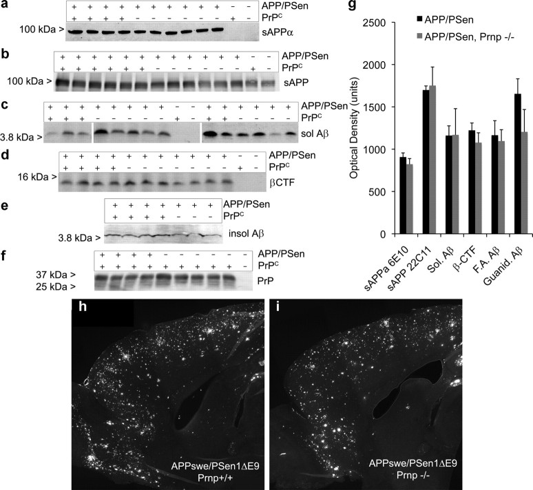
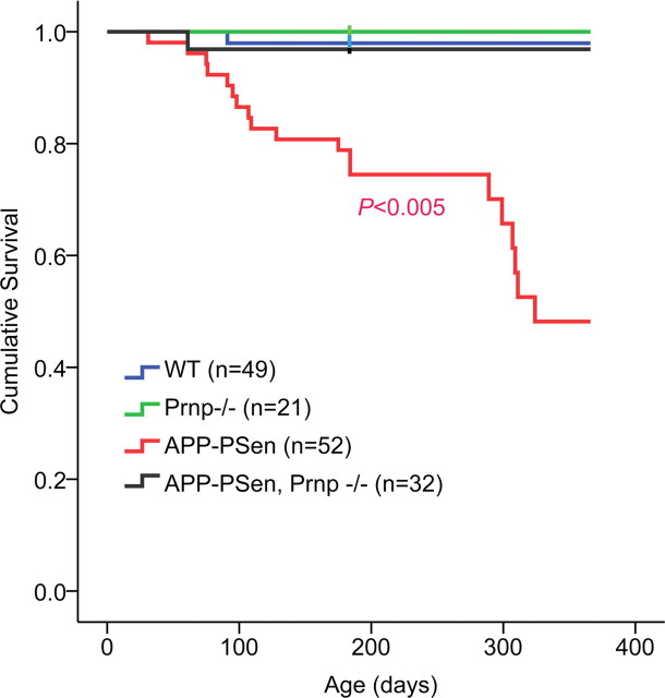
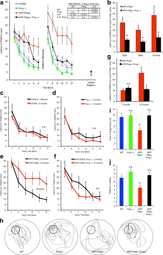
Soluble oligomers of the amyloid-beta (Abeta) peptide are thought to play a key role in the pathophysiology of Alzheimer's disease (AD). Recently, we reported that synthetic Abeta oligomers bind to cellular prion protein (PrP(C)) and that this interaction is required for suppression of synaptic plasticity in hippocampal slices by oligomeric Abeta peptide. We hypothesized that PrP(C) is essential for the ability of brain-derived Abeta to suppress cognitive function. Here, we crossed familial AD transgenes encoding APPswe and PSen1DeltaE9 into Prnp-/- mice to examine the necessity of PrP(C) for AD-related phenotypes. Neither APP expression nor Abeta level is altered by PrP(C) absence in this transgenic AD model, and astrogliosis is unchanged. However, deletion of PrP(C) expression rescues 5-HT axonal degeneration, loss of synaptic markers, and early death in APPswe/PSen1DeltaE9 transgenic mice. The AD transgenic mice with intact PrP(C) expression exhibit deficits in spatial learning and memory. Mice lacking PrP(C), but containing Abeta plaque derived from APPswe/PSen1DeltaE9 transgenes, show no detectable impairment of spatial learning and memory. Thus, deletion of PrP(C) expression dissociates Abeta accumulation from behavioral impairment in these AD mice, with the cognitive deficits selectively requiring PrP(C).


Figures

Figure 1.
Figure 1.
APP and Aβ levels are not dependent on PrPC. a–f, Immunoblot analysis of soluble brain extracts (a–c, f) or detergent-extracted particulate fractions (d) or guanidinium-solubilized material (e) with anti-Aβ 6E10 antibody (a, c–e), anti-APP 22C11 antibody (b), or anti-PrPC 6D11 antibody (f). Genotype is indicated at the top of each blot for separate mice. The migration of MW standards is shown at the left. g, Densitometric analysis of immunoblot signals from experiments as in a–f. Data are mean ± SEM for n = 5–12 mice per genotype. h, i, Immunofluorescent detection of Aβ in a parasagittal section of the anterior half of a APPswe/PSen1ΔE9 transgenic mouse brain at 12 months of age of the Prnp+/+ (h) or Prnp−/− (i) genotype.
Figure 2.
Figure 2.
Serotonin axons and synaptic markers are preserved in APPswe/PSen1ΔE9 mice lacking PrPC. a, Anti-GFAP immunohistology of parasagittal brain sections was scored for astrocytic area in the anterior half of the cerebral cortex. Data are mean ± SEM for n = 10 mice per genotype. b, Parasagittal sections of cerebral cortex from mice of the indicated genotypes were stained with anti-5-HT. Projections of 30 μm confocal Z-stacks are shown. Note the reduced fiber length in the APPswe/PSen1ΔE9 sample. Scale bar, 50 μm. c, The 5-HT-immunoreactive fiber length in the parasagittal plane from images as in b was measured. Data are mean ± SEM for n = 10–16 mice per genotype. *p < 0.05, one-way ANOVA with post hoc comparisons as indicated; n.s., no significant difference. d, Cerebral cortex from mice of the indicated genotypes was stained with anti-synaptophysin antibody and imaged by confocal microscopy with a 100× objective. Scale bar, 10 μm. e, f, The fractional area of immunoreactive puncta from images as in d is reported as a function of genotype. Sections were stained with anti-synaptophysin (e) or anti-PSD-95 (f). Data are mean ± SEM for n = 4–8 sites from each of 9–10 mice per genotype. *p < 0.05, one-way ANOVA with post hoc comparisons.
Figure 3.
Figure 3.
Survival of APP-PSen mice improved by absence of PrPC. Survival of wild-type and APPswe/PSen1ΔE9 transgenic mice with and without PrPC expression. Survival of the APPswe/PSen1ΔE9 mice is significantly shorter than each of the other three genotypes by the Wilcoxon statistic (p < 0.005).
Figure 4.
Figure 4.
Deletion of PrPC expression rescues spatial learning and memory in APPswe-PSen1ΔE9 transgenic mice. a, Spatial learning is plotted as the latency to find a hidden platform in the Morris water maze at 12 months of age. After the first set of swim trials, the platform location was reversed for the second set of trials. At a later time, after the probe trial, latency to reach a visible platform was scored. Data are mean ± SEM for these groups: C57BL/6, n = 19 (8 male, 11 female); Prnp−/− n = 15 (7 male, 8 female); APP-PSen n = 11 (6 male, 5 female); APP-PSen, Prnp−/− n = 20 (11 male, 9 female). Performance differed by genotype across trial blocks 5–6 and 9–12 (two-way RM-ANOVA: APP-PSen by Prnp interaction, p = 0.015; APP-PSen, p < 0.001; Prnp, p < 0.001). For the indicated Tukey post hoc pairwise comparisons, the APP-PSen group differed from each of the other three groups (p < 0.001), but none of the other genotypes differed significantly from one another. b, The performance of the APPswe/PSen1ΔE9 transgenic mice with and without PrPC expression from trial blocks 11–12 of a is separated by sex. The latencies are reduced in the Prnp−/− groups compared to the Prnp+/+ groups, *p < 0.05, two-tailed Student's t test. c–f, Mice of each genotype were tested for spatial learning at 3 months, and the data are compared to the 12 month performance curves replotted from a. By RM-ANOVA, only the APP-PSen group differed significantly (p < 0.05) between age groups. Data are mean ± SEM for n = 11–20 mice per group. g, The performance of the APPswe/PSen1ΔE9 transgenic mice with and without PrPC expression from trial blocks 11–12 of d–f is separated by age. The latencies are reduced in the Prnp−/− groups compared to the Prnp+/+ at 12 months but not 3 months, *p < 0.05, two-tailed Student's t test. h, Two days after the second set of hidden platform learning trials from a, a 60 s probe trial was conducted. The swim track of one representative mouse from each of the four genotypes is illustrated. A small circle indicates the learned platform location. i, j, The time spent at the site of the previous platform location (i) and crosses of the previous platform location (j) are shown. Data are mean ± SEM for n = 11–20 mice per group. *p < 0.05, one-way ANOVA with post hoc comparison to the WT group.

Comment in

Similar articles

Cited by

References

    1. Austin MC, Whitehead RE, Edgar CL, Janosky JE, Lewis DA. Localized decrease in serotonin transporter-immunoreactive axons in the prefrontal cortex of depressed subjects committing suicide. Neuroscience. 2002;114:807–815. - PubMed
    1. Balducci C, Beeg M, Stravalaci M, Bastone A, Sclip A, Biasini E, Tapella L, Colombo L, Manzoni C, Borsello T, Chiesa R, Gobbi M, Salmona M, Forloni G. Synthetic amyloid-beta oligomers impair long-term memory independently of cellular prion protein. Proc Natl Acad Sci U S A. 2010;107:2295–2300. - PMC - PubMed
    1. Baumann F, Tolnay M, Brabeck C, Pahnke J, Kloz U, Niemann HH, Heikenwalder M, Rülicke T, Bürkle A, Aguzzi A. Lethal recessive myelin toxicity of prion protein lacking its central domain. EMBO J. 2007;26:538–547. - PMC - PubMed
    1. Berlyne DE. Curiosity and exploration. Science. 1966;153:25–33. - PubMed
    1. Berlyne DE, Koenig ID, Hirota T. Novelty, arousal, and the reinforcement of diversive exploration in the rat. J Comp Physiol Psychol. 1966;62:222–226. - PubMed

Publication types

MeSH terms