. 2010 May 5;30(18):6434-42.
doi: 10.1523/JNEUROSCI.4066-09.2010.

Local control of postinhibitory rebound spiking in CA1 pyramidal neuron dendrites

Affiliations

Local control of postinhibitory rebound spiking in CA1 pyramidal neuron dendrites

Giorgio A Ascoli et al. J Neurosci. .

Abstract

Postinhibitory rebound spiking is characteristic of several neuron types and brain regions, where it sustains spontaneous activity and central pattern generation. However, rebound spikes are rarely observed in the principal cells of the hippocampus under physiological conditions. We report that CA1 pyramidal neurons support rebound spikes mediated by hyperpolarization-activated inward current (I(h)), and normally masked by A-type potassium channels (K(A)). In both experiments and computational models, K(A) blockage or reduction consistently resulted in a somatic action potential upon release from hyperpolarizing injections in the soma or main apical dendrite. Rebound spiking was systematically abolished by the additional blockage or reduction of I(h). Since the density of both K(A) and I(h) increases in these cells with the distance from the soma, such "latent" mechanism may be most effective in the distal dendrites, which are targeted by a variety of GABAergic interneurons. Detailed computer simulations, validated against the experimental data, demonstrate that rebound spiking can result from activation of distal inhibitory synapses. In particular, partial K(A) reduction confined to one or few branches of the apical tuft may be sufficient to elicit a local spike following a train of synaptic inhibition. Moreover, the spatial extent and amount of K(A) reduction determines whether the dendritic spike propagates to the soma. These data suggest that the plastic regulation of K(A) can provide a dynamic switch to unmask postinhibitory spiking in CA1 pyramidal neurons. This newly discovered local modulation of postinhibitory spiking further increases the signal processing power of the CA1 synaptic microcircuitry.


Figures

Figure 1.
Figure 1.
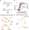
Postinhibitory rebound spiking in CA1 pyramidal neurons: hypothesis and experimental validation. a, Simulated (left) and experimental (right) response to somatic hyperpolarization in control conditions, after blocking KA, and after blocking both KA and Ih. b, Dual recordings of dendritic and somatic membrane potential obtained during a dendritic current injection (−1 nA, 200 ms at ∼250 μm from the soma). The drawing on the left shows the experimental configuration. c, Voltage traces of the dendritic membrane potential recorded in response to a train (20 IPSCs at 100 Hz) injected in the apical trunk at 300 μm from the soma in control conditions, in the presence of Ba2+ (150 μm), and in the presence of Ba2+ and ZD 7288 (20 μm). The current trace below represents the injected train of IPSCs. d, Somatic voltage traces recorded in response to a 200 ms, −400 pA current injection to show the differences in sag and rebound from a neuron that shows rebound spiking (black trace) and one that does not (gray trace). e, Cumulative probability to have a rebound spike as a function of the time constant of the sag; symbols on the two sides of the curve show the average values (±SEM) for the two groups of cells, those that show rebound firing (black) and those that do not (gray); the inset shows the number of neurons initiating a rebound spike as a function of the amplitude of somatic current injection.
Figure 2.
Figure 2.
Postinhibitory rebound spiking in CA1 pyramidal neurons: computational model. a, Drawings of the two model neurons used in all simulations (scale bar: 100 μm). b, Dendritic distribution for KA (closed circles, same distribution for both neurons) and Ih (triangles). c, Simultaneous fitting of both somatic and dendritic experimental recordings in control conditions, performed independently for the two model neurons. Actual dendritic locations for current injection were at 219 and 224 μm from the soma for neurons pc2b and ri06, respectively. d, Model traces for neuron pc2b, using the same experimental protocol described in Figure 1b.
Figure 3.
Figure 3.
Dissecting the mechanism underlying rebound spiking in CA1 pyramidal cells. a, Dendritic membrane potential from neuron pc2b, during a dendritic current injection (−1 nA, 200 ms) under control conditions (black trace), with a 50% higher input resistance (pink), with reduced KA (10% of control, orange), and with Ih with a halved activation/deactivation time constant and reduced KA (10% of control, green trace). b, Left, Somatic voltage traces recorded in response to a 200 ms, 500 pA hyperpolarizing current injection, under control conditions (black), in the presence of 2 mm 4-AP (red), and in the presence of 4-AP and 20 μm ZD 7288 (blue); right, somatic voltage traces from simulations with the model including KM, with (bottom, green) or without (top, red) KD. c, Plot of normalized KA activation in the subthreshold range of membrane potential.
Figure 4.
Figure 4.
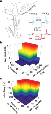
Local modulation of KA can result in a dendritic rebound spike that may or not propagate to the soma. a, Typical results obtained stimulating two different stretches of dendritic membrane (blue, 280 μm2; red, 368 μm2) in the same neuron (pc2b), with the same number of synapses (n = 25, schematically indicated on the red branch). In both cases, somatic and dendritic membrane potentials (traces on the right) are plotted after different reduction (15% or 40% of control) of local KA channels. b, Conditions to initiate a dendritic rebound spike in the blue branch with different combinations of the peak synaptic conductance, Ih somatic density, and synaptic train duration after reducing the local KA to 40% of control. Equal colors represent combinations with the same peak synaptic conductance. c, Conditions required to initiate a dendritic rebound spike in the blue or in the red branch with different combinations of the stimulation frequency, Ih time constant, and decay time constant of the inhibitory synapses. Equal colors represent combinations with the same stimulation frequency; in both branches, synapses were activated 20 times, with the local KA reduced to 15% of control.
Figure 5.
Figure 5.
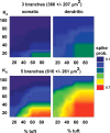
Average dendritic or somatic spike probability as a function of local KA channel density (% of control) and amount of the tuft on which KA is reduced. Synaptic stimulation was localized in contiguous branches of different sizes. The average spike probability was calculated by testing all combinations of three (top panels) and five (bottom panels) branches in neuron pc2b.

References

    1. Anderson AE, Adams JP, Qian Y, Cook RG, Pfaffinger PJ, Sweatt JD. Kv4.2 phosphorylation by cyclic AMP-dependent protein kinase. J Biol Chem. 2000;275:5337–5346. - PubMed
    1. Andrásfalvy BK, Mody I. Differences between the scaling of miniature IPSCs and EPSCs recorded in the dendrites of CA1 mouse pyramidal neurons. J Physiol. 2006;576:191–196. - PMC - PubMed
    1. Armstrong WE, Stern JE. Phenotypic and state-dependent expression of the electrical and morphological properties of oxytocin and vasopressin neurones. Prog Brain Res. 1998;119:101–113. - PubMed
    1. Ascoli GA, Donohue DE, Halavi M. NeuroMorpho.Org: a central resource for neuronal morphologies. J Neurosci. 2007;27:9247–9251. - PMC - PubMed
    1. Balu R, Strowbridge BW. Opposing inward and outward conductances regulate rebound discharges in olfactory mitral cells. J Neurophysiol. 2007;97:1959–1968. - PubMed

Publication types

MeSH terms

Substances

LinkOut - more resources