. 2010 Sep;24(9):3298-309.
doi: 10.1096/fj.09-147488. Epub 2010 May 5.

Formation of oral and pharyngeal dentition in teleosts depends on differential recruitment of retinoic acid signaling

Affiliations

Formation of oral and pharyngeal dentition in teleosts depends on differential recruitment of retinoic acid signaling

Yann Gibert et al. FASEB J. 2010 Sep.

Abstract

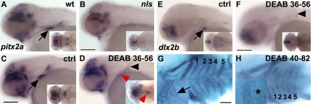
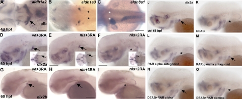
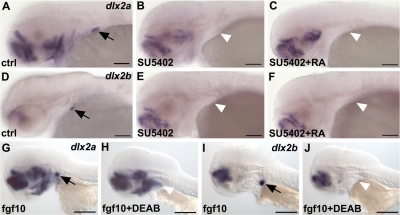
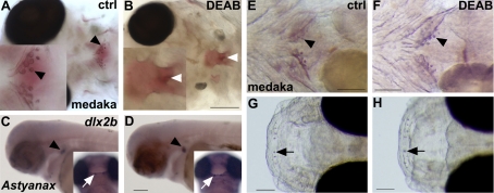
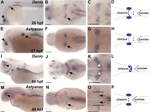
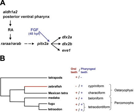
One of the goals of evolutionary developmental biology is to link specific adaptations to changes in developmental pathways. The dentition of cypriniform fishes, which in contrast to many other teleost fish species possess pharyngeal teeth but lack oral teeth, provides a suitable model to study the development of feeding adaptations. Here, we have examined the involvement of retinoic acid (RA) in tooth development and show that RA is specifically required to induce the pharyngeal tooth developmental program in zebrafish. Perturbation of RA signaling at this stage abolished tooth induction without affecting the development of tooth-associated ceratobranchial bones. We show that this inductive event is dependent on RA synthesis from aldh1a2 in the ventral posterior pharynx. Fibroblast growth factor (FGF) signaling has been shown to be critical for tooth induction in zebrafish, and its loss has been associated with oral tooth loss in cypriniform fishes. Pharmacological treatments targeting the RA and FGF pathways revealed that both pathways act independently during tooth induction. In contrast, we find that in Mexican tetra and medaka, species that also possess oral teeth, both oral and pharyngeal teeth are induced independently of RA. Our analyses suggest an evolutionary scenario in which the gene network controlling tooth development obtained RA dependency in the lineage leading to the cypriniforms. The loss of pharyngeal teeth in this group was cancelled out through a shift in aldh1a2 expression, while oral teeth might have been lost ultimately due to deficient RA signaling in the oral cavity.


Figures

Figure 1.
Figure 1.
RA is required for pharyngeal tooth induction. A, B) pitx2a expression is absent in the ventral posterior pharynx in nls/aldh1a2 at 56 hpf (B) as compared with wild types (WT; A). C–H) Wild-type embryos treated with DEAB from 36 hpf (CF) or 40 hpf (G, H) onward fixed at 56 hpf (CF) or 82 hpf (G, H). In DEAB-treated embryos, pitx2a is faintly detected in the pharyngeal epithelium (D, red arrowhead) and is located in a group of cells at the midline (D, inset, red arrowhead). dlx2b expression is not detected in tooth buds in DEAB-treated embryos at 56 hpf (F). Alcian blue staining at 82 hpf of a control embryo (G) shows all branchial arches numbered from 1 to 5, including teeth (G, inset). In DEAB-treated embryos, all ceratobranchial arches are present (H), but teeth are absent. Asterisk marks the absence of tooth. Black arrowheads denote the presence of the pectoral fin that is present when DEAB is applied at late stage (later than 13 hpf; ref. 36). Scale bars = 100 μm.
Figure 2.
Figure 2.
RA, provided by aldh1a2, serves as a signal for tooth induction that is transduced by raraa receptors. A–C) Expression of the zebrafish aldh1a2, aldh1a3, and aldh8a1 genes at 43 hpf in dorsal views. A) aldh1a2 is mainly detected in the dorsal spinal cord, the pectoral fin mesenchyme, and 2 patches of cells in the ventral posterior pharynx engulfing the fifth ceratobranchial arch (arrow). B) aldh1a3 is expressed in the otic vesicle. C) aldh8a1 is expressed in the liver and the anterior gut. Asterisks in B, C mark the location where teeth develop. D–I) nls embryos exposed to exogenous RA during gastrulation and somitogenesis fail to restore tooth induction (F, I). Asterisk in F marks lack of teeth. +3 RA indicates that exogenous RA was added during gastrulation, somitogenesis and for tooth induction. +2 RA indicates that exogenous RA was added during gastrulation and somitogenesis only. All in situ hybridizations were performed at 60 hpf. J–O) Tooth induction is mediated by raraa subtypes. Inhibition of rara subtypes blocks tooth induction, as marked by absence of dlx2a expression (L), while activating raras in embryos lacking RA signaling restores tooth induction (N). The rarg subtypes play no role during tooth induction (M, O). Asterisks in K, L, O denote the absence of tooth bud expression (pfb, pectoral fin bud). Scale bars = 100 μm.
Figure 3.
Figure 3.
RA and FGF signaling act independently during tooth induction. A–F) Exogenous RA applied at 32 hpf does not restore dlx2a (C) or dlx2b (F) expression when Fgf signaling has been blocked at 32 hpf. G–J) Ectopic activation of FGF signaling using an inducible fgf10a construct does not restore dlx2a (H) or dlx2b (J) expression in RA-depleted embryos. White arrowheads indicate absence of tooth induction. All in situ hybridizations were performed at 56 hpf. Scale bars = 100 μm.
Figure 4.
Figure 4.
RA is not required for oral and pharyngeal tooth induction in noncypriniform teleosts. A, B) Alizarin red staining of medaka embryos treated with DEAB during somitogenesis shows lack of pharyngeal teeth (white arrowhead in B) and the most posterior ceratobranchial arch (B), confirming the potency of DEAB in these species (ventral views). C, D) A. mexicanus control embryo (C) and DEAB-treated embryo from 30 hpf onward (D), showing dlx2b expression in oral (white arrow) and pharyngeal (black arrowhead) teeth (lateral views). E–H) Alizarin red staining of medaka embryos treated with DEAB from 3–7 dpf shows pharyngeal (F, arrowhead) and upper oral teeth (H, arrow; lateral views). Scale bars = 100 μm.
Figure 5.
Figure 5.
aldh1a2 expression in zebrafish and Mexican tetra embryos. A–H) aldh1a2 expression in embryos at 26 hpf for zebrafish and 27 hpf for Mexian tetra. Arrows indicate expression in the caudal branchial arch; arrowheads denote expression in the dorsal retina. I–P) aldh1a2 expression at 44 hpf. Arrows indicate the 2 patches of cells in the ventral posterior pharynx in zebrafish (J) and the expression in the caudal branchial arch in Astyanax (N). White lines (K, O) demarcate the midline of the embryo; black lines (O) the limit of aldh1a2 expression in the pharynx toward the midline. Asterisks (D, H, K, L, P) mark the putative location of the induction of the first tooth. Note that this location is within the aldh1a2-positive cells for the zebrafish (K) but not for the Astyanax as marked by the black arrows (O). Scale bars = 100 μm.
Figure 6.
Figure 6.
Proposed model of genetic pathways involved in pharyngeal tooth induction in zebrafish. A) aldh1a2 from the posterior ventral pharynx generates RA. This signal activates the RAR subtype α. RA signaling is required for a proper expression of pitx2a; however, in absence of this signal, pitx2a is still detected at a lower level as a spot at the midline. Dotted lines represent an RA requirement for proper expression of pitx2a. pitx2a, in turn, regulates the expression of dlx2a, dlx2b, and eve1. FGF signaling is required after RA signaling (48 vs. 43 hpf). Moreover we know from previous study that a lack of FGF signaling does not abolish pitx2a expression in the tooth bud . B) Phylogenetic tree of the main fish models showing the presence of absence of a particular set of teeth (oral vs. pharyngeal). Red branch represents the hijack of RA for tooth induction in zebrafish.

Similar articles

Cited by

References

    1. Smith M M, Fraser G J, Mitsiadis T A. Dental lamina as source of odontogenic stem cells: evolutionary origins and developmental control of tooth generation in gnathostomes. J Exp Zoolog B Mol Dev Evol. 2009;312B:260–280. - PubMed
    1. Miletich I, Sharpe P T. Neural crest contribution to mammalian tooth formation. Birth Defects Res C Embryo Today. 2004;72:200–212. - PubMed
    1. Mitsiadis T A, Smith M M. How do genes make teeth to order through development? J Exp Zoolog B Mol Dev Evol. 2006;306:177–182. - PubMed
    1. Tompkins K. Molecular mechanisms of cytodifferentiation in mammalian tooth development. Connect Tissue Res. 2006;47:111–118. - PubMed
    1. Stock D W. Zebrafish dentition in comparative context. J Exp Zoolog B Mol Dev Evol. 2007;308:523–549. - PubMed

Publication types

MeSH terms

Associated data

LinkOut - more resources