. 2010 Oct;51(10):4913-20.
doi: 10.1167/iovs.09-4892. Epub 2010 May 5.

Retinal pigment epithelium defects accelerate photoreceptor degeneration in cell type-specific knockout mouse models of choroideremia

Affiliations

Retinal pigment epithelium defects accelerate photoreceptor degeneration in cell type-specific knockout mouse models of choroideremia

Tanya Tolmachova et al. Invest Ophthalmol Vis Sci. 2010 Oct.

Abstract

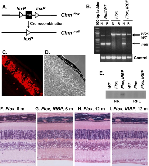
Purpose: Choroideremia (CHM) is a progressive X-linked degeneration of three ocular layers (photoreceptors, retinal pigment epithelium, and choroid), with a complex and still largely unclear pathogenesis. To investigate the pathophysiology of CHM, the authors engineered mice with a cell type-specific Chm/Rep1 knockout (KO).

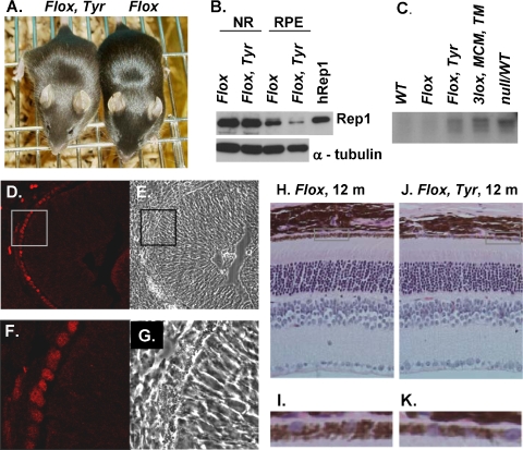
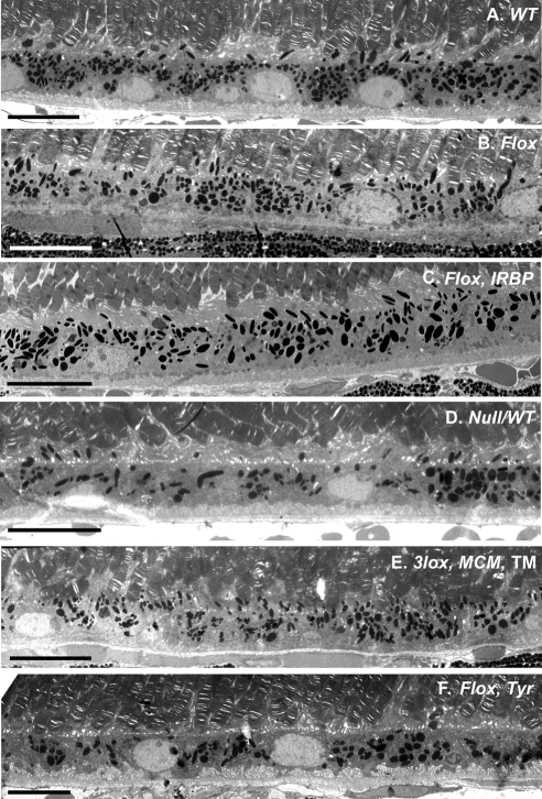
Methods: A mouse line carrying a conditional allele Chm(Flox) was crossed with the transgenic line IRBP-Cre to achieve Chm KO, specifically in the photoreceptor layer, and Tyr-Cre to produce Chm KO, specifically in the retinal pigment epithelial and other pigmented cells. Chm(Flox), Tyr-Cre+ and Chm(Flox), IRBP-Cre+ mice were mated to produce mice with Chm KO in both layers. All mouse lines were studied by histology, electron microscopy, electroretinography (ERG), scanning laser ophthalmoscopy (SLO), and biochemical

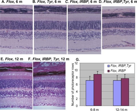
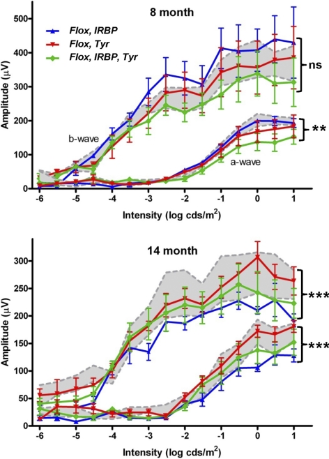
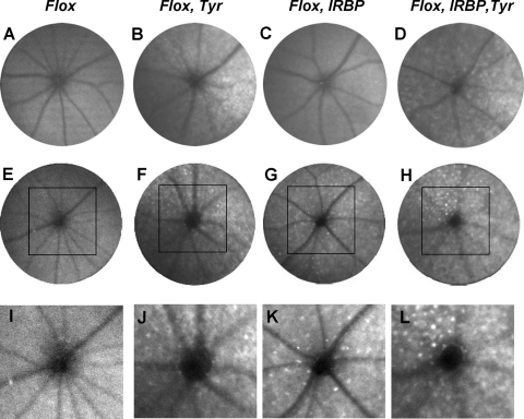
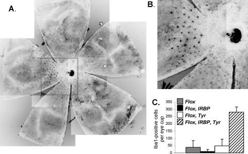
Results: In Chm(Flox), IRBP-Cre+ mice the authors observed the progressive degeneration of photoreceptors in the presence of normal retinal pigment epithelium (RPE). Chm(Flox), Tyr-Cre+ mice exhibited coat color dilution and pigment abnormalities of the RPE in the presence of an intact outer nuclear layer. In 6- to 8-month-old Chm(Flox), Tyr-Cre+, IRBP-Cre+ mice, the degeneration of photoreceptors was accelerated compared with Chm(Flox), IRBP-Cre+ mice but became leveled with age, such that it was comparable at 12 to 14 months. Detailed ERG and SLO analysis supported the histopathologic findings.

Conclusions: Defects in photoreceptors and RPE can arise because of intrinsic defects caused cell autonomously by the Chm KO. However, when both photoreceptors and RPE are diseased, the dynamics of the degenerative process are altered. Photoreceptor functional deficit and cell death manifest much earlier, suggesting that the diseased RPE accelerates photoreceptor degeneration.


Figures

Figure 1.
Figure 1.
Characterization of ChmFlox, IRBP-Cre+ model. (A) Illustration showing main features of the ChmFlox allele and its conversion to the ChmNull allele after Cre-recombination. (B) Neuroretina (N) and RPE (R) were isolated from the eyes of ChmNull/WT, ChmFlox, ChmFlox, IRBP-Cre+ animals and analyzed by PCR using primers that could identify ChmNull, ChmFlox, ChmWT alleles (top) and control primers (bottom). (C) Eyes were collected from ChmWT, IRBP-Cre+ animals at P21, stained with Cre-specific primary antibody and Alexa-568 secondary antibody, and visualized by confocal microscopy. (D) Phase image corresponding to (C). (E) In vitro prenylation reaction was performed on the cytosolic fractions of the lysates isolated from neuroretina (NR) and the RPE of the ChmWT, ChmFlox, and ChmFlox, IRBP-Cre+ animals. (F–I) Paraffin wax sections were prepared from the eyes of ChmFlox animals at 6 months (F) and 12 months (H) and ChmFlox, IRBP-Cre+ animals at 6 months (G) and 12 months (I), cut at 4-μm thickness, and stained with hematoxylin and eosin.
Figure 2.
Figure 2.
Characterization of ChmFlox, Tyr-Cre+ model (A) Photograph of 4-week-old ChmFlox, Tyr-Cre+ and ChmFlox littermates. (B) Immunoblot analysis of the neuroretina (NR) and RPE cells isolated from the eyes of the ChmFlox and ChmFlox, Tyr-Cre+ animals using Rep-specific antibody J905 and α-tubulin antibody (as a loading control). (C) In vitro prenylation reaction was performed on the cytosolic fractions of the lysates isolated from the RPE of the ChmWT, ChmFlox, ChmNull/WT, TM-induced ChmFlox, MerCreMer+ and ChmFlox, Tyr-Cre+ animals. (D–G) Frozen section were prepared from ChmWT, Tyr-Cre+ animals at E12.5, stained with Cre-specific primary antibody and Alexa-568 secondary antibody, and visualized by confocal microscopy. (E) Phase image corresponding to (D). (F) Enlarged area of the inset in (D). (G) Phase image corresponding to (F). (H–K) Paraffin wax sections were prepared from the eyes of ChmFlox (H) and ChmFlox, Tyr-Cre+ animal (J) at 1 year, cut at 4-μm thickness, and stained with hematoxylin and eosin. (I) Enlargement of the boxed area in (H). (K) Enlargement of the boxed area in (J).
Figure 3.
Figure 3.
Electron microscopy of the ChmWT (A), ChmFlox (B), ChmFlox, IRBP-Cre+ (C), Chm Null/WT (D), TM -induced Chm3lox, MerCreMer+ (E), ChmFlox, Tyr-Cre+ (F) at 5 months. In (A), (B), (C), and (E), the RPE is highly pigmented, and melanosomes are found in apical processes. In contrast, in (F) and some areas of (D), pigmentation is altered and melanosomes remain in the cell body. Scale bar, 10 μm.
Figure 4.
Figure 4.
Histologic analysis of the retina of the 6-month old ChmFlox (A), ChmFlox, Tyr-Cre+ (B), ChmFlox, IRBP-Cre+ (C), ChmFlox, IRBP-Cre+, Tyr-Cre+ (D) and 12-month old ChmFlox (E), IRBP-Cre+, Tyr-Cre+ (F). (G) Morphometric analysis of the number of photoreceptor rows in the central retina in the ChmFlox, IRBP-Cre+ (purple bars) and ChmFlox, IRBP-Cre+, Tyr-Cre+ (blue bars). Numbers of animals analyzed were: ChmFlox, IRBP-Cre+: 6 to 8 months (n = 15), 12 to 14 months (n = 19); ChmFlox, IRBP-Cre+, Tyr-Cre+: 6 to 8 months (n = 7), 12 to 14 months (n = 11).
Figure 5.
Figure 5.
Quantification of the amplitude of the major components (a- and b-wave) of the dark-adapted ERG across a range of stimulus intensity. Two groups of animals (8-month- and 14-month-old) were studied. Numbers of 8-month-old animals were: ChmFlox, Tyr-Cre+ (n = 5); ChmFlox, IRBP-Cre+ (n = 3); ChmFlox, Tyr-Cre+, IRBP-Cre+ (n = 5), and ChmFlox (n = 4). Numbers of 14-month-old animals were: ChmFlox, Tyr-Cre+ (n = 6); ChmFlox, IRBP-Cre+ (n = 5); ChmFlox, Tyr-Cre+, IRBP-Cre+ (n = 6), and ChmFlox (n = 6). All genotypes are shown on each graph: ChmFlox, Tyr-Cre+ (red line), ChmFlox, IRBP-Cre+ (blue line), ChmFlox, IRBP-Cre+, Tyr-Cre+ (green line). Mean amplitudes ± SEM are plotted. Values for control animals (ChmFlox, mean ± SEM) are indicated by gray shading.
Figure 6.
Figure 6.
Representative AF-SLO projection fundus images obtained from ChmFlox (A, E, I), ChmFlox, Tyr-Cre+ (B, F, J), ChmFlox, IRBP-Cre+ (C, G, K), ChmFlox, IRBP-Cre+, Tyr-Cre+ (D, H, L) at 8 months (A–D) and 14 months (E–L). (I–L) Enlargement of the boxed area in (E–H).
Figure 7.
Figure 7.
(A) Retinal flat mounts from an 11-month-old ChmFlox IRBP-Cre+, Tyr-Cre+ animal were stained with anti–Iba1 primary and anti–rabbit Alexa-488 secondary antibody. (B) Enlargement of the boxed area in (A). (C) Quantitative data of the number of Iba1-positive cells per eye cup from 11- to 12-month-old ChmFlox (gray bar), ChmFlox, IRBP-Cre+ (closed bar), ChmFlox, Tyr-Cre+ (open bar), and ChmFlox, IRBP-Cre+, Tyr-Cre+ (hatched bar) animals.

References

    1. Cremers FPM, Ropers H. Choroideremia. In: Scriver CR, Beaudet AL, Sly WS, et al., eds. The Metabolic and Molecular Bases of Inherited Disease. New York: McGraw-Hill, Inc.; 2001:5935–5945
    1. Heckenlively JR, Bird AJ. Choroideremia. In: Heckenlively JR. ed. Retinitis Pigmentosa. Philadelphia: JB Lippincott; 1988:176–187
    1. McCulloch C. Choroideremia and other choroidal atrophies. In: Newsome DA. ed. Retinal Dystrophies and Degenerations. New York: Raven Press; 1988:285–295
    1. MacDonald IM, Sereda C, McTaggart K, Mah D. Choroideremia gene testing. Expert Rev Mol Diagn. 2004;4:478–484 - PubMed
    1. Yau RJ, Sereda CA, McTaggart KE, Sauve Y, MacDonald IM. Choroideremia carriers maintain a normal electro-oculogram (EOG). Doc Ophthalmol. 2007;114:147–151 - PubMed

Publication types

Substances