

Cerebellar atrophy in human and murine succinic semialdehyde dehydrogenase deficiency
- PMID: 20445195
- PMCID: PMC3155424
- DOI: 10.1177/0883073810368137
Cerebellar atrophy in human and murine succinic semialdehyde dehydrogenase deficiency
Abstract
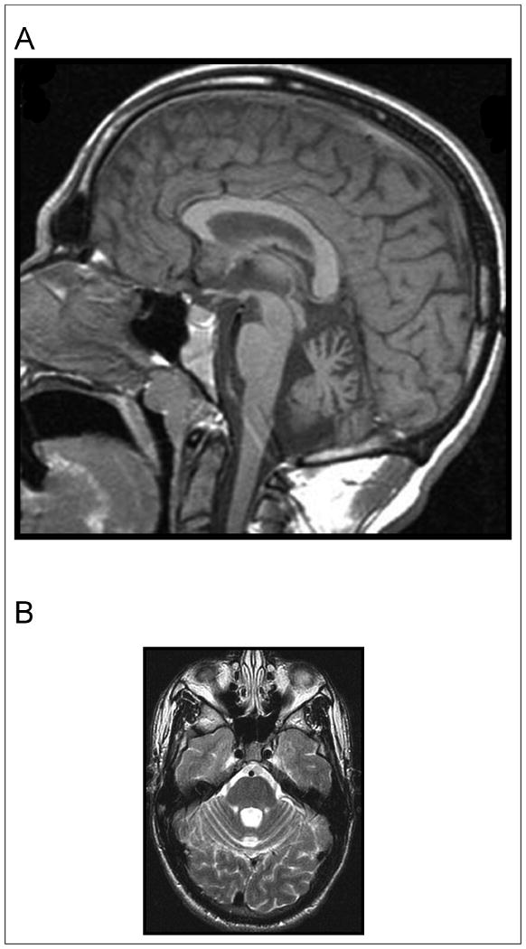
Human succinic semialdehyde dehydrogenase deficiency, an autosomal recessive disorder of γ-aminobutyric acid (GABA) catabolism, was modeled by a murine model sharing the phenotype of ataxia and seizures. Magnetic resonance imaging (MRI) with volumetry was obtained on 7 patients versus controls, and MRI with stereology was derived in 3 murine genotypes: null, wild-type, and heterozygous mutants. All patients had T1 hypointensity and T2 hyperintensity in globus pallidus, and 5 also had similar changes in subthalamic and cerebellar dentate nuclei. There was a trend for patients to have a smaller cerebellar vermis. Homozygous null mice had significantly lower total brain and cerebellar volumes than wild-types and heterozygotes. Stereology confirmed cerebellar atrophy and was otherwise normal in multiple regions. Cerebellar volume loss is present in the murine disorder with a trend for cerebellar atrophy in patients. Reduced cerebellar volume can reflect neurodegeneration and may be related to the clinical manifestations.
Conflict of interest statement
Figures
References
-
- Chambliss KL, Caudle DL, Hinson DD, et al. Molecular cloning of the mature NAD(+)-dependent succinic semialdehyde dehydrogenase from rat and human. cDNA isolation, evolutionary homology, and tissue expression. J Biol Chem. 1995;270:461–467. - PubMed
-
- Gibson KM, Aramaki S, Sweetman L, et al. Stable isotope dilution analysis of 4-hydroxybutyric acid: an accurate method for quantification in physiological fluids and the prenatal diagnosis of 4-hydroxybutyric aciduria. Biomed Environ Mass Spectrom. 1990;19:89–93. - PubMed
-
- Pearl PL, Gibson KM, Acosta MT, et al. Clinical spectrum of succinic semialdehyde dehydrogenase deficiency. Neurology. 2003;60:1413–1417. - PubMed
-
- Pearl PL, Acosta MT, Wallis DD, et al. Dyskinetic features of succinate semialdehyde dehydrogenase deficiency, a GABA degradative defect. In: Fernandez-Alvarez E, Arzimanoglu A, Tolosa E, editors. Paediatric Movement Disorders: Progress in Understanding. Montrouge, France: John Libbey Eurotext; 2005. pp. 203–212.
