. 2010 Aug;130(8):2017-30.
doi: 10.1038/jid.2010.108. Epub 2010 May 6.

PKC-delta and -eta, MEKK-1, MEK-6, MEK-3, and p38-delta are essential mediators of the response of normal human epidermal keratinocytes to differentiating agents

Affiliations

PKC-delta and -eta, MEKK-1, MEK-6, MEK-3, and p38-delta are essential mediators of the response of normal human epidermal keratinocytes to differentiating agents

Gautam Adhikary et al. J Invest Dermatol. 2010 Aug.

Erratum in

Abstract

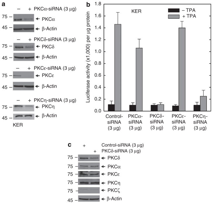
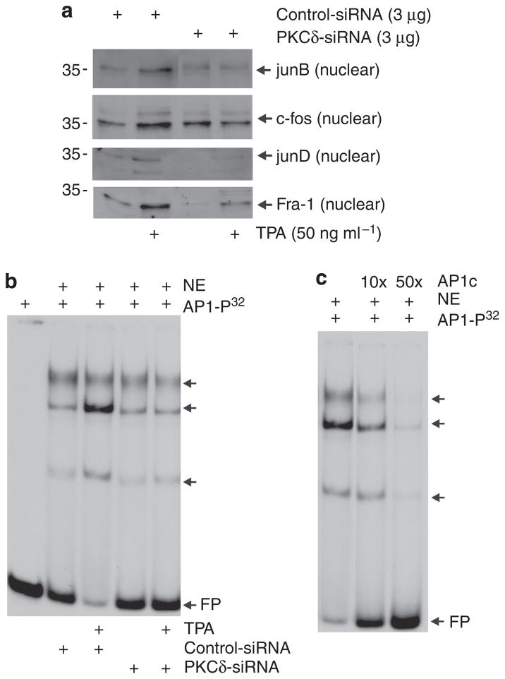
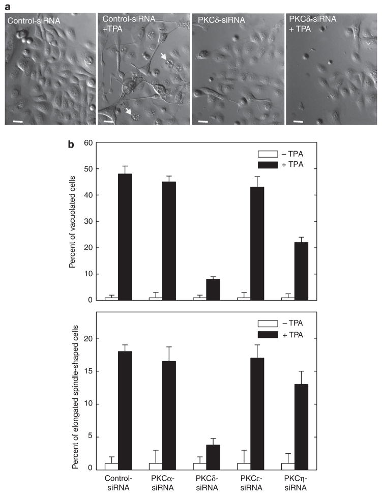
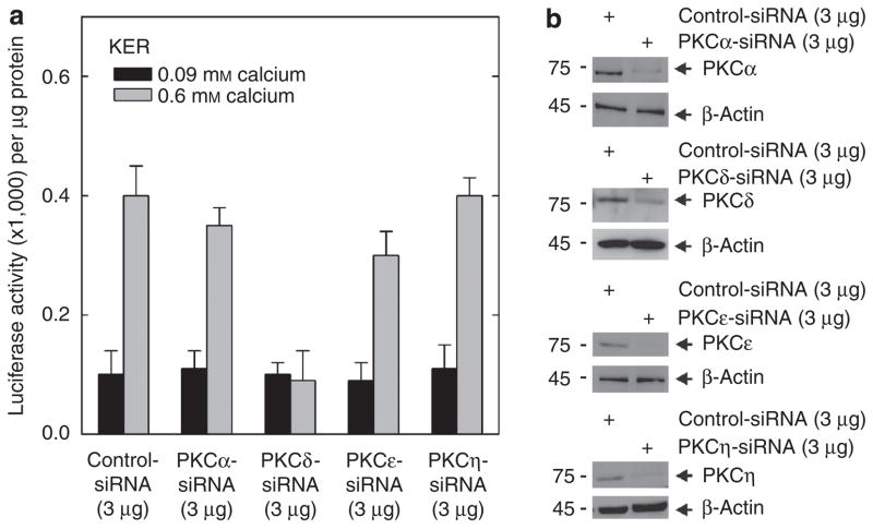
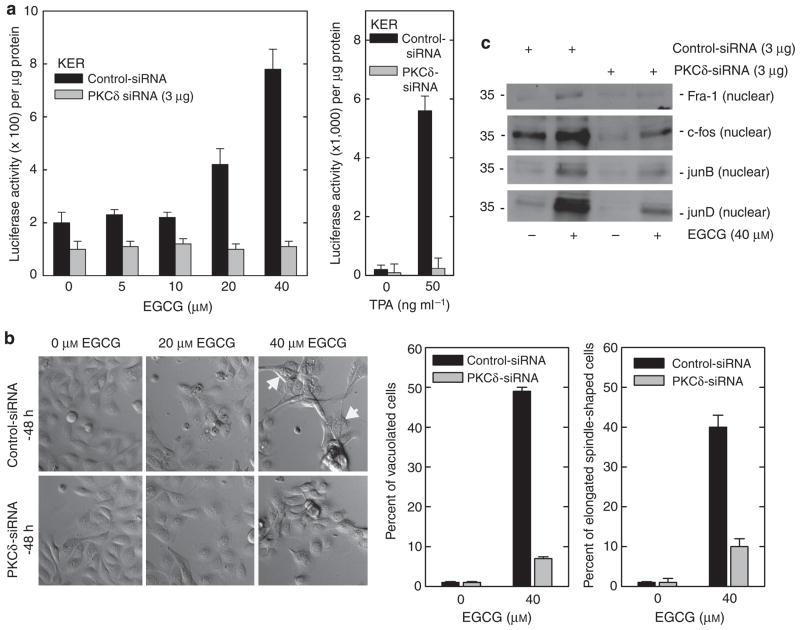
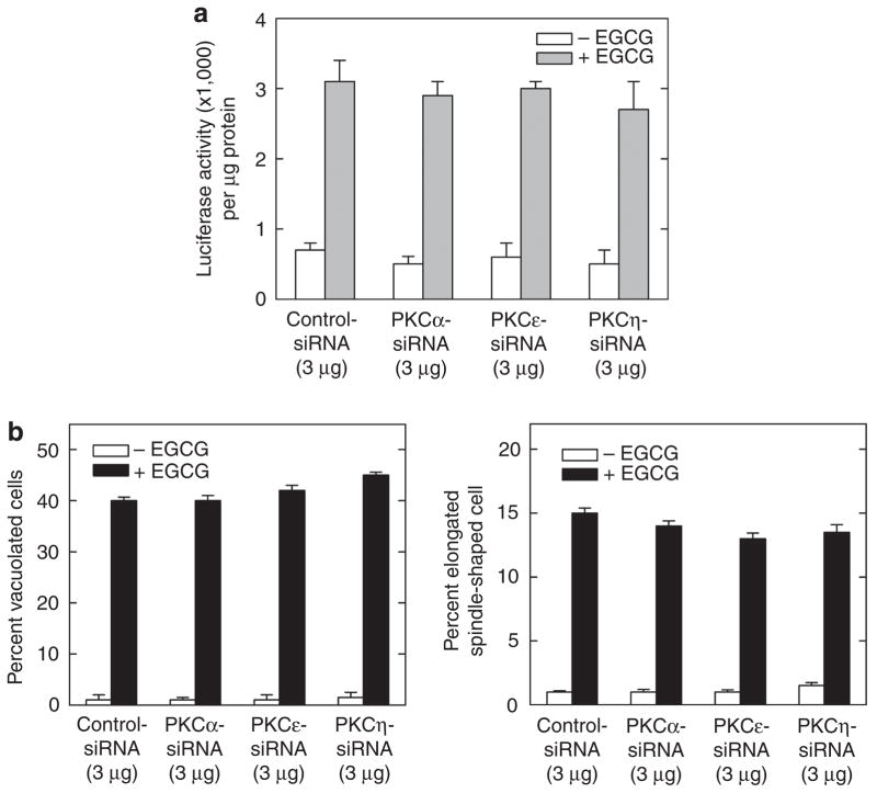
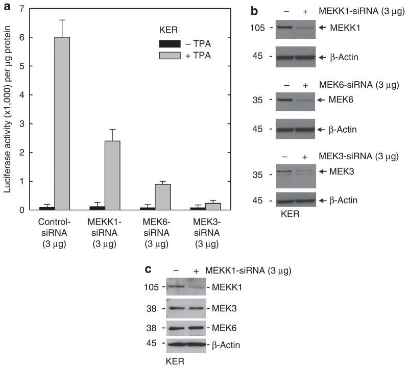
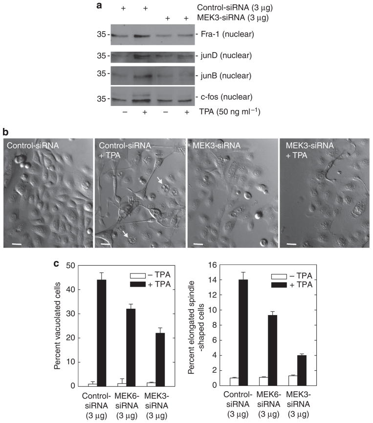
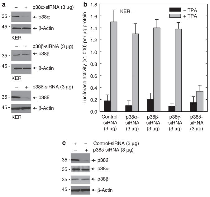
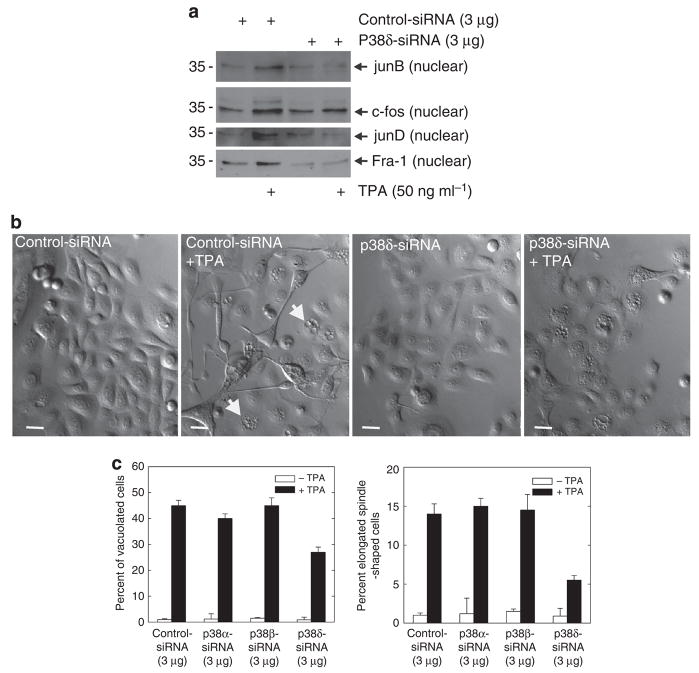
Previous studies suggest that the novel protein kinase C (PKC) isoforms initiate a mitogen-activated protein kinase (MAPK) signaling cascade that regulates keratinocyte differentiation. However, assigning these functions has relied on treatment with pharmacologic inhibitors and/or manipulating kinase function using overexpression of wild-type or dominant-negative kinases. As these methods are not highly specific, an obligatory regulatory role for individual kinases has not been assigned. In this study, we use small interfering RNA knockdown to study the role of individual PKC isoforms as regulators of keratinocyte differentiation induced by the potent differentiating stimulus, 12-O-tetradecanoylphorbol-13-acetate (TPA). PKC-delta knockdown reduces TPA-activated involucrin promoter activity, nuclear activator protein-1 factor accumulation and binding to DNA, and cell morphology change. Knockdown of PKC downstream targets, including MEKK-1, MEK-6, MEK-3, or p38-delta, indicates that these kinases are required for these responses. Additional studies indicate that knockdown of PKC-eta inhibits TPA-dependent involucrin promoter activation. In contrast, knockdown of PKC-alpha (a classical PKC isoform) or PKC-epsilon (a novel isoform) does not inhibit these TPA-dependent responses. Further studies indicate that PKC-delta is required for calcium and green tea polyphenol-dependent regulation of end responses. These findings are informative as they suggest an essential role for selected PKC and MAPK cascade enzymes in mediating a range of end responses to a range of differentiation stimuli in keratinocytes.


Conflict of interest statement

CONFLICT OF INTEREST

The authors state no conflict of interest.

Figures

Figure 1
Figure 1. PKC-δ and PKC-η are required for the TPA-dependent hINV promoter activation
(a) Knockdown of PKC isoform expression. Keratinocytes were electroporated with 3 μg of control siRNA or siRNA specifying the indicated PKC isoforms per 1 × 10⁶ cells. Total cell extract was prepared at 48 hours after electroporation and 25 μg of protein was electrophoresed for immunodetection of each PKC isoform. For purposes of convenience, we routinely measured PKC levels at 48 hours after application of the siRNA; however, the levels remain reduced for up to 4 days (not shown). This experiment was repeated four times and the siRNA-dependent reduction ranged as follows: PKC-α, 50–70%; PKC-δ, 50–70%; PKC-ε, 80–90%, and PKC-η, 70–90%, as measured by densitometry. (b) PKC-δ is required for TPA-dependent promoter activation. At 48 hours after siRNA electroporation, cells were electroporated with 3 μg of endotoxin-free pINV-2473. After an additional 6 hours, the cells were treated with 50 ng ml−1 TPA and luciferase activity was assessed 18 hours later. The error bars represent the mean±SD of a representative experiment. Similar results were observed in each of four separate experiments. The t-test analysis reveals that PKC-δ and PKC-η siRNAs significantly suppress (P<0.01) TPA-stimulated promoter activity when compared with control siRNA, n = 4. The lesser reduction associated with PKC-α siRNA treatment was not significant (P<0.98). (c) PKC-δ knockdown does not influence the level of other PKC isoforms. Keratinocytes were electroporated with 3 μg of control siRNA or PKC-δ siRNA per 1 × 10⁶ cells. Total cell extract was prepared at 48 hours after electroporation and 25 μg of protein was electrophoresed for immunodetection of each PKC isoform. hINV, human involucrin; PKC, protein kinase C; siRNA, small interfering RNA; TPA, 12-O-tetradecanoylphorbol-13-acetate.
Figure 2
Figure 2. PKC-δ knockdown reduces TPA-dependent nuclear accumulation of AP-1 factors
Keratinocytes were electroporated with 3 μg of control or PKC-δ siRNA per 1 × 10⁶ cells. After 24 hours, cells were treated with 0 or 50 ng ml−1 TPA for an additional 48 hours before preparation of nuclear extracts. (a) Nuclear extract (5 μg) was electrophoresed for immunological detection of junB, c-fos, junD, and Fra-1. (b) Reduced PKC-δ level is associated with reduced TPA-induced AP-1 factor DNA binding. Electrophoretic mobility shift assay was performed as outlined in the Materials and Methods. The arrows indicate bands that bind to 32P-AP-1 double-stranded oligonucleotide. (c) Nuclear extracts from cells treated with control siRNA and 50 ng ml−1 TPA were incubated with 32P-AP-1 in the presence of 10- or 50-fold excess of radioinert AP-1 double-stranded oligonucleotide before non-denaturing gel electrophoresis. The arrows indicate migration of the gel-shifted bands and FP indicates migration of the free probe. AP-1, activator protein-1; Fra-1, Fos-related antigen-1; FP, migration of free probe; NE, nuclear extract; PKC, protein kinase C; siRNA, small interfering RNA; TPA, 12-O-tetradecanoylphorbol-13-acetate.
Figure 3
Figure 3. PKC-δ knockdown reduces TPA effect on cell morphology
(a) Keratinocytes were electroporated with 3 μg of control or PKC-δ siRNA per 1 × 10⁶ cells. After 24 hours, cells were treated with 0 or 50 ng ml−1 TPA for an additional 48 hours before monitoring of cell morphology. Cells expressing normal PKC-δ level seem vacuolated and take on a spindle-shaped morphology, and this response is attenuated in PKC-δ siRNA-treated cells. The arrows indicate vacuolated cells. Bars = 25 μm in length. Phase-contrast images were obtained using an Olympus IX81 motorized inverted microscope (Hamburg, Germany) and a × 20 objective. (b) Keratinocytes were electroporated with 3 μg siRNA encoding the indicated PKC isoform and treated with TPA as indicated above, and after an additional 48 hours morphology was assessed. The plots were generated by determining the percentage of cells containing large intracellular vacuoles or showing an elongated spindle shape (see photographs) out of a minimum of 100 cells on each of three slides. The results are expressed as the mean±SD. This experiment is representative of four repeated experiments. The t-test analysis reveals that PKC-δ and PKC-η siRNAs significantly suppress (P<0.01) TPA-stimulated cell shape change when compared with control siRNA, n = 4. PKC, protein kinase C; siRNA, small interfering RNA; TPA, 12-O-tetradecanoylphorbol-13-acetate.
Figure 4
Figure 4. PKC-δ is required for the calcium-associated increase in hINV promoter activity
(a) Keratinocytes were electroporated with 3 μg of control siRNA or the indicated PKC-specific siRNA per 1 × 10⁶ cells. At 2 days after siRNA electroporation, the cells were trypsinized, collected by centrifugation, and electroporated with 3 μg of endotoxin-free hINV promoter-luciferase reporter plasmid. After 6 hours, the cells were treated with KSFM (0.09 mM calcium) or KSFM containing 0.6 mM calcium. After an additional 18 hours, the cells were harvested and extracts were assayed for luciferase activity. The results are expressed as the mean±SD. This experiment is representative of three repeated experiments. The t-test analysis reveals that PKC-δ siRNA significantly suppresses (P<0.01) calcium-stimulated promoter activity below the activity observed with control siRNA, n = 3. (b) Keratinocytes were treated with siRNA as indicated above and PKC isoform level was monitored after 48 hours. The experiment was repeated three times and the siRNA-dependent reduction in PKC isoform level ranged as follows: PKC-α, 50–70%; PKC-δ, 50–70%; PKC-ε, 80–90%, and PKC-η, 70–90%. hINV, human involucrin; KSFM, keratinocyte serum-free medium; PKC, protein kinase C; siRNA, small interfering RNA.
Figure 5
Figure 5. PKC-δ is required for the EGCG-dependent hINV promoter activity, nuclear AP-1 factor accumulation, and morphology change
(a) Keratinocytes were electroporated with 3 μg of control or PKC-δ specific siRNA per 1 × 10⁶ cells. At 48 hours after siRNA electroporation, cells were electroporated with 3 μg of endotoxin-free pINV-2473. After an additional 6 hours, the cells were treated with 0–40 μM EGCG (left panel) or 50 ng ml−1 TPA (right panel) and luciferase activity was assessed 18 hours later. The results are expressed as the mean±SD. The t-test analysis reveals that PKC-δ siRNA significantly suppresses 20 and 40 μM EGCG-dependent promoter activity (P<0.01) below the activity observed with control siRNA, n = 3. (b) PKC-δ is required for EGCG-dependent morphological change. Cells expressing normal PKC-δ levels show a spindle-like shape and accumulate intracellular vacuoles, a response that is attenuated in PKC-δ-knockdown cells. The plots were generated by determining the percentage of cells containing large intracellular vacuoles or showing an elongated spindle shape (see photographs) out of a minimum of 100 cells on each of three slides. The results are expressed as the mean±SD. This experiment is representative of three repeated experiments. PKC-δ siRNA significantly suppresses (Po0.01) EGCG-stimulated cell shape change when compared with control siRNA, n = 3. Bar = 25 μm. (c) PKC-δ is required for EGCG-dependent nuclear accumulation of AP-1 factors. Nuclear extract (5 μg), prepared from cells treated with 3 mg of control or PKC-δ siRNA for 48 hours, was electrophoresed and nuclear levels of Fra-1, c-fos, junB, and junD were detected by immunoblot. AP-1, activator protein-1; EGCG, (-)-epigallocatechin-3-gallate; Fra-1, Fos-related antigen-1; PKC, protein kinase C; siRNA, small interfering RNA; TPA, 12-O-tetradecanoylphorbol-13-acetate.
Figure 6
Figure 6. PKCs α, ε, and η are not required for EGCG-dependent hINV promoter activity or morphology change
(a) PKC α, ε, and η isoforms are not required for EGCG-stimulated hINV promoter activity. Keratinocytes were electroporated with 3 μg of control or PKC-δ-specific siRNA per 1 × 10⁶ cells. At 48 hours after siRNA electroporation, cells were electroporated with 3 μg of endotoxin-free pINV-2473. After an additional 6 hours, the cells were treated with 40 μM EGCG and luciferase activity was assessed 18 hours later. The results are expressed as the mean±SD. This experiment is representative of four repeated experiments. (b) PKC α, ε, and η isoforms are not required for EGCG-dependent morphology change. Keratinocytes were electroporated with siRNA encoding the indicated PKC isoform and treated with EGCG for 48 hours as indicated above and morphology was assessed. The plots were generated by determining the percentage of cells containing large intracellular vacuoles or showing an elongated spindle-shaped morphology out of a minimum of 100 cells on each of three slides. The results are expressed as the mean±SD. This experiment is representative of four repeated experiments. EGCG, (-)-epigallocatechin-3-gallate; hINV, human involucrin; PKC, protein kinase C; siRNA, small interfering RNA; TPA, 12-O-tetradecanoylphorbol-13-acetate.
Figure 7
Figure 7. MEKK-1, MEK-6, and MEK-3 are required for TPA-dependent hINV promoter activation
(a) Keratinocytes were electroporated with 3 μg of control siRNA or siRNA encoding MEKK-1, MEK-6, or MEK-3 per 1 × 10⁶ cells. After 48 hours, cells were electroporated with 3 μg of endotoxin-free pINV-2473. After another 6 hours, the cells were treated with 50 ng ml−1 TPA and luciferase activity was assessed after 18 hours. The error bars represent the mean±SD of a representative experiment. The t-test analysis reveals that MEKK-1 (P<0.05), MEK-6 (P<0.01), and MEK-3 (P<0.01) siRNAs significantly suppress TPA-stimulated promoter activity when compared with control siRNA, n = 3. (b) Reduced MEKK-1, MEK-6, and MEK-3 levels. Total extract (25 μg) was prepared from cells 48 hours after treatment with siRNA and the indicated proteins were detected by immunoblot. This experiment was repeated four times and the siRNA-dependent reduction in level ranged as follows: MEKK-1, 70–90%; MEK-6, 70–90%; and MEK-3 70–90%, as measured by densitometry. (c) MEKK1 siRNA does not influence the levels of other MAPK cascade kinases. Keratinocytes were electroporated with 3 μg of control siRNA or siRNA encoding the MEKK-1 per 1 × 10⁶ cells. After 48 hours, cells were harvested and the level of the indicated protein was monitored. Similar results were observed in each of three experiments. hINV, human involucrin; MEK, MAPK/ERK kinase; MEKK-1, MAPK kinase kinase-1; PKC, protein kinase C; siRNA, small interfering RNA; TPA, 12-O-tetradecanoylphorbol-13-acetate.
Figure 8
Figure 8. MEK-3 knockdown reduces TPA-dependent nuclear AP-1 factor accumulation and morphological response
Keratinocytes were electroporated with 3 mg of control or MEK-3 siRNA per 1 × 10⁶ cells. After 24 hours, cells were treated with 50 ng ml−1 TPA for an additional 48 hours. (a) Nuclear extract (5 μg) was electrophoresed for detection of junB, c-fos, junD, and Fra-1. (b) MEK-3 siRNA reduces the effect of TPA treatment on keratinocyte morphology. Keratinocytes were electroporated with 3 μg of MEK-3 siRNA and then treated for an additional 48 hours with TPA before assessment of morphology. Bars = 25 μm. The arrows indicate cells containing large intracellular vacuoles. (c) Effect of MEK-6 and MEK-3 knockdown on TPA-induced keratinocyte morphology change. Keratinocytes were electroporated with 3 μg of specific siRNA and then treated with 50 ng ml−1 TPA for 48 hours before assessing the effect on morphology. The plots were generated by determining the percentage of cells containing large intracellular vacuoles or showing an elongated spindle shape (see pictures) out of a minimum of 100 cells on each of three slides. The results are expressed as the mean±SD. MEK-6 (P<0.05) and MEK-3 (P<0.01) siRNAs significantly suppress TPA-stimulated morphology change when compared with control siRNA, n = 3. AP-1, activator protein-1; Fra-1, Fos-related antigen-1; MEK, MAPK/ERK kinase; siRNA, small interfering RNA; TPA, 12-O-tetradecanoylphorbol-13-acetate.
Figure 9
Figure 9. p38-δ is required for the TPA-dependent hINV promoter activation
(a) Knockdown of p38 isoform expression. Keratinocytes were electroporated with 3 μg of control siRNA or siRNA encoding the indicated p38 isoforms per 1 × 10⁶ cells. Total cell extract was prepared at 48 hours after electroporation and 25 μg protein was electrophoresed for immunodetection of each p38 isoform. This experiment was repeated four times and the siRNA-dependent reduction in level ranged as follows: p38-α, 80–90%; p38-β, 70–90%; and p38-δ, 80–90%, as measured by densitometry. (b) p38-δ is required for TPA-dependent hINV promoter activation. At 48 hours after siRNA electroporation, cells were electroporated with 3 μg of endotoxin-free pINV-2473. After an additional 6 hours, the cells were treated with 50 ng ml−1 TPA and luciferase activity was assessed 18 hours later. The results are expressed as the mean±SD. p38-δ siRNA significantly suppresses (P<0.01) TPA-stimulated promoter activity below the activity observed with control siRNA, n = 3. (c) p38-δ knockdown does not influence the level of other p38 isoforms. Keratinocytes were electroporated with 3 μg of control or p38-δ isoform-specific siRNA per 1 × 10⁶ cells. Total cell extract was prepared at 48 hours after electroporation and 25 μg of protein was electrophoresed for immunodetection of each p38 isoform. This experiment was repeated twice with similar results. hINV, human involucrin; siRNA, small interfering RNA; TPA, 12-O-tetradecanoylphorbol-13-acetate.
Figure 10
Figure 10. p38-δ knockdown affects nuclear AP-1 factor level and cell morphology
(a) p38-δ is required for TPA-dependent nuclear localization of AP-1 factors. Keratinocytes were electroporated with 3 mg of control or p38-δ siRNA per 1 × 10⁶ cells. After 24 hours, cells were treated with 50 ng ml−1 TPA for an additional 48 hours. Nuclear extract (5 μg) was electrophoresed for detection of junB, c-fos, junD, and Fra-1. (b) p38-δ knockdown reduces the effect of TPA on keratinocyte morphology. Keratinocytes were electroporated with 3 mg of control or p38-δ siRNA per 1 × 10⁶ cells. After 24 hours, cells were treated with 50 ng ml−1 TPA for an additional 48 hours. Bars = 25 μm. The arrows indicate vacuolated cells. (c) p38-α and p38-β knockdown does not reduce the effect of TPA on keratinocyte morphology. Keratinocytes were treated with siRNA and TPA as indicated above before assessment of cell morphology. The plots were generated by determining the percentage of cells containing large intracellular vacuoles or showing an elongated spindle-shaped morphology out of a minimum of 100 cells on each of three slides. The results are expressed as the mean±SD. p38-δ siRNA significantly suppresses (P<0.01) TPA-stimulated morphology change below the activity observed with control siRNA, n = 3. AP-1, activator protein-1; Fra-1, Fos-related antigen-1; siRNA, small interfering RNA; TPA, 12-O-tetradecanoylphorbol-13-acetate.

Comment in

References

    1. Acs P, Beheshti M, Szallasi Z, et al. Effect of a tyrosine 155 to phenylalanine mutation of protein kinase cdelta on the proliferative and tumorigenic properties of NIH 3T3 fibroblasts. Carcinogenesis. 2000;21:887–91. - PubMed
    1. Alt A, Ohba M, Li L, et al. Protein kinase Cdelta-mediated phosphorylation of alpha6beta4 is associated with reduced integrin localization to the hemidesmosome and decreased keratinocyte attachment. Cancer Res. 2001;61:4591–8. - PubMed
    1. Amar LS, Shabana A, Oboeuf M, et al. Desmosomes are regulated by protein kinase C in primary rat epithelial cells. Cell Adhes Commun. 1998;5:1–12. - PubMed
    1. Ashendel CL. The phorbol ester receptor: a phospholipid-regulated protein kinase. Biochim Biophys Acta. 1985;822:219–42. - PubMed
    1. Aziz MH, Sundling KE, Dreckschmidt NE, et al. Protein kinase Cepsilon inhibits UVR-induced expression of FADD, an adaptor protein, linked to both Fas- and TNFR1-mediated apoptosis. J Invest Dermatol. 2009;129:2011–21. - PMC - PubMed

Publication types

MeSH terms

Substances