. 2010:2010:250978.
doi: 10.4061/2010/250978. Epub 2010 Mar 22.

Histological and immunohistochemical evaluation of autologous cultured bone marrow mesenchymal stem cells and bone marrow mononucleated cells in collagenase-induced tendinitis of equine superficial digital flexor tendon

Affiliations

Histological and immunohistochemical evaluation of autologous cultured bone marrow mesenchymal stem cells and bone marrow mononucleated cells in collagenase-induced tendinitis of equine superficial digital flexor tendon

Antonio Crovace et al. Vet Med Int. 2010.

Abstract

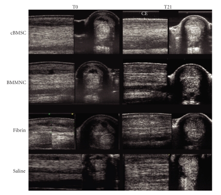
The aim of this study was to compare treatment with cultured bone marrow stromal cells (cBMSCs), bone marrow Mononucleated Cells (BMMNCs), and placebo to repair collagenase-induced tendinitis in horses. In six adult Standardbred horses, 4000 IU of collagenase were injected in the superficial digital flexor tendon (SDFT). Three weeks after collagenase treatment, an average of either 5.5 x 10(6) cBMSCs or 1.2 x 10(8) BMMNCs, fibrin glue, and saline solution was injected intralesionally in random order. In cBMSC- and BMMNCS-treated tendons, a high expression of cartilage oligomeric matrix protein (COMP) and type I collagen, but low levels of type III collagen were revealed by immunohistochemistry, with a normal longitudinally oriented fiber pattern. Placebo-treated tendons expressed very low quantities of COMP and type I collagen but large numbers of randomly oriented type III collagen fibers. Both cBMSC and BMMNCS grafts resulted in a qualitatively similar heling improvement of tendon extracellular matrix, in terms of the type I/III collagen ratio, fiber orientation, and COMP expression.


Figures

Figure 1
Figure 1
Time course description of the study performed over a period of 24 weeks (T-3-T21); US data were recorded immediately prior to collagenase injection to induce tendon injury, recorded as T-3, then 3 weeks later, just prior to injection of either cBMSC or BMMNCS suspension, fibrin glue, or placebo (= saline) into the tendon lesion (T0), and again at 3, 6, 8, 12, 16, 18, and 21 weeks (T3-T21) following tendon treatment. At T21, animals were euthanized and the SDFTs of all four limbs were harvested and processed for histology and immunohistochemistry examination.
Figure 2
Figure 2
Longitudinal and transverse ultrasound images recorded on T0, that is, the day of injection of intralesional cultured bone marrow mesenchymal cells (cBMSC), bone marrow Mononucleated Cells (BMMNCS), fibrin and saline, and at T21, 21 weeks after treatment.
Figure 3
Figure 3
Tissue slides obtained from SDFTs injected with cBMSCs, and BMMNCS. Tissue stains with hematoxylin-eosin (H and E) and Herovyci (Hero) highlight the longitudinal orientation of collagen fibers after transplantation of cBMSCs and BMMNCS; Histological examination of the sham tendons, both with HE and Herovyci staining, showed the presence of mature collagen (i.e., predominantly strong red Herovyci staining of fibers in tendon sections) and a longitudinal fiber orientation, signifying a normal tendon architecture. Additionally, the absence or very scanty mononuclear cell infiltrate was observed in all normal control tendons. No inflammatory or other reactions to the fibrin glue were observed in any of the treated limbs, and at T21 no fibrin residues were recognized in microscopic examinations. (T21) Tissue slides from the superficial digital flexor tendons (SDFT) following experimentally induced tissue injury and subsequent treatment with cultured bone marrow mesenchymal stem cells (cBMSCs (a)) and Bone Marrow Mononucleated Cells (BMMNCS (b)). Incomplete maturation of collagen was verified by the observation of blue staining fibers mixed with red ones. In BMMNCS a large number of mononucleated cells are recognizable in the interfascicular zone. Immunohistochemistry stains for Collagen type I (C/I) and collagen type III (C/III) show a high expression of C/I and a very low expression of C/III. Stains for cartilage oligomeric matrix protein (COMP) expression. BMMNCS-treated SDFT (b) show a strong, diffuse expression like sham SDFT (Figure 4(c)). Immunohistochemistry staining for the expression of CD34+ mononucleated cells in cBMSCs (a) shows a low expression limited to the interfascicular zone. In BMMNCS (b)-treated SDFT, the presence of numerous mononucleated CD34+ cells near microvessels, which sometimes show some CD34+ endotheliocytes in their wall, which is highlighted. In cBMSC- and BMMNCS-treated SDFTs, microscopic examination at T21 revealed a longitudinal orientation of newly formed collagen fibers. At this time point, sections stained by the Herovyci method revealed minimal areas of blue-stained thin, well-oriented precollagen fibers mixed with well-differentiated and- oriented red-stained mature collagen fibers, indicating a still somewhat incomplete collagen maturation at this time in the treatment. In BMMNCS-treated SDFTs also, large numbers of mononucleated cells were present in the interfascicular zone. In contrast, in placebo SDFTs, injected with saline or fibrin, the tissue architecture was disrupted or markedly modified, showing blue precollagen and red collagen fibers randomly or not longitudinally oriented, and a relatively loss of the architectural pattern.
Figure 3
Figure 3
Tissue slides obtained from SDFTs injected with cBMSCs, and BMMNCS. Tissue stains with hematoxylin-eosin (H and E) and Herovyci (Hero) highlight the longitudinal orientation of collagen fibers after transplantation of cBMSCs and BMMNCS; Histological examination of the sham tendons, both with HE and Herovyci staining, showed the presence of mature collagen (i.e., predominantly strong red Herovyci staining of fibers in tendon sections) and a longitudinal fiber orientation, signifying a normal tendon architecture. Additionally, the absence or very scanty mononuclear cell infiltrate was observed in all normal control tendons. No inflammatory or other reactions to the fibrin glue were observed in any of the treated limbs, and at T21 no fibrin residues were recognized in microscopic examinations. (T21) Tissue slides from the superficial digital flexor tendons (SDFT) following experimentally induced tissue injury and subsequent treatment with cultured bone marrow mesenchymal stem cells (cBMSCs (a)) and Bone Marrow Mononucleated Cells (BMMNCS (b)). Incomplete maturation of collagen was verified by the observation of blue staining fibers mixed with red ones. In BMMNCS a large number of mononucleated cells are recognizable in the interfascicular zone. Immunohistochemistry stains for Collagen type I (C/I) and collagen type III (C/III) show a high expression of C/I and a very low expression of C/III. Stains for cartilage oligomeric matrix protein (COMP) expression. BMMNCS-treated SDFT (b) show a strong, diffuse expression like sham SDFT (Figure 4(c)). Immunohistochemistry staining for the expression of CD34+ mononucleated cells in cBMSCs (a) shows a low expression limited to the interfascicular zone. In BMMNCS (b)-treated SDFT, the presence of numerous mononucleated CD34+ cells near microvessels, which sometimes show some CD34+ endotheliocytes in their wall, which is highlighted. In cBMSC- and BMMNCS-treated SDFTs, microscopic examination at T21 revealed a longitudinal orientation of newly formed collagen fibers. At this time point, sections stained by the Herovyci method revealed minimal areas of blue-stained thin, well-oriented precollagen fibers mixed with well-differentiated and- oriented red-stained mature collagen fibers, indicating a still somewhat incomplete collagen maturation at this time in the treatment. In BMMNCS-treated SDFTs also, large numbers of mononucleated cells were present in the interfascicular zone. In contrast, in placebo SDFTs, injected with saline or fibrin, the tissue architecture was disrupted or markedly modified, showing blue precollagen and red collagen fibers randomly or not longitudinally oriented, and a relatively loss of the architectural pattern.
Figure 4
Figure 4
(T21) Tissue slides from the superficial digital flexor tendons (SDFT) following experimentally induced tissue injury and subsequent treatment with Fibrin (c), Saline (d), and normal tendon (e). Tissue stains with hematoxylin-eosin (H and E) and Herovyci (Hero) show loss of the longitudinal fiber pattern and crimp in tendons treated with-Fibrin (c); in Saline treated tendon (d), there is complete disruption with randomly oriented fibers, clearly shown in the central area. The collagen I/III ratio in Fibrin treated tendon (c) is clearly lower than in normal (e) and cell grafted tendon (Figures 3(a) and 3(b)); in Saline (d) treated tendon, the central zone was highly positive for collagen type III but not for type I. Stains for cartilage oligomeric matrix protein (COMP) show stack and spread expression in both control-treated SDFT (c-d). In immunohistochemistry staining for the expression of CD34+ mononucleated cells in Fibrin (c), Saline (d) and Normal (e) Tendon, only sporadic CD34+ stained mononuclear elements were detected. By contrast, in all three placebo-treated SDFTs, the location of the lesion was readily identified as an area with high positivity for type III collagen, very low expression of type I collagen and in some zones, evidence of mineralization. Similarly to the trend of type I collagen expression, COMP was expressed in a homogeneous and diffuse pattern in sham untreated control SDFTs as well as in cBMSC- and BMMNCS-injected tendons. Scant, weak expression of COMP, restricted to areas that circumscribed the injected portions of the tendon, was observed in all three placebo-treated SDFTs. As expected, no CD34+ mononuclear cells were observed in the sham tendon sections. Similar results were obtained in sections of placebo-injected SDFTs, although a certain number of inflammatory cells were present, interspersed in perivascular areas of these tendons (data not shown). Only sporadic CD34+ stained mononuclear elements (i.e., 0 to 2 per HPF) were observed in cBMSC-injected tendons (data not shown). On the contrary, in BMMNCS-treated SDFTs large numbers of mononucleated cells, in part CD34+ stained, were present in the interfascicular zone.
Figure 4
Figure 4
(T21) Tissue slides from the superficial digital flexor tendons (SDFT) following experimentally induced tissue injury and subsequent treatment with Fibrin (c), Saline (d), and normal tendon (e). Tissue stains with hematoxylin-eosin (H and E) and Herovyci (Hero) show loss of the longitudinal fiber pattern and crimp in tendons treated with-Fibrin (c); in Saline treated tendon (d), there is complete disruption with randomly oriented fibers, clearly shown in the central area. The collagen I/III ratio in Fibrin treated tendon (c) is clearly lower than in normal (e) and cell grafted tendon (Figures 3(a) and 3(b)); in Saline (d) treated tendon, the central zone was highly positive for collagen type III but not for type I. Stains for cartilage oligomeric matrix protein (COMP) show stack and spread expression in both control-treated SDFT (c-d). In immunohistochemistry staining for the expression of CD34+ mononucleated cells in Fibrin (c), Saline (d) and Normal (e) Tendon, only sporadic CD34+ stained mononuclear elements were detected. By contrast, in all three placebo-treated SDFTs, the location of the lesion was readily identified as an area with high positivity for type III collagen, very low expression of type I collagen and in some zones, evidence of mineralization. Similarly to the trend of type I collagen expression, COMP was expressed in a homogeneous and diffuse pattern in sham untreated control SDFTs as well as in cBMSC- and BMMNCS-injected tendons. Scant, weak expression of COMP, restricted to areas that circumscribed the injected portions of the tendon, was observed in all three placebo-treated SDFTs. As expected, no CD34+ mononuclear cells were observed in the sham tendon sections. Similar results were obtained in sections of placebo-injected SDFTs, although a certain number of inflammatory cells were present, interspersed in perivascular areas of these tendons (data not shown). Only sporadic CD34+ stained mononuclear elements (i.e., 0 to 2 per HPF) were observed in cBMSC-injected tendons (data not shown). On the contrary, in BMMNCS-treated SDFTs large numbers of mononucleated cells, in part CD34+ stained, were present in the interfascicular zone.

References

    1. Awad HA, Butler DL, Boivin GP, et al. Autologous mesenchymal stem cell-mediated repair of tendon. Tissue Engineering. 1999;5(3):267–277. - PubMed
    1. Ouyang HW, Goh JC, Thambyah A, et al. Knitted poly-lactide-co-glycolide scaffold loaded with bone marrow stromal cells in repair and regeneration of rabbit achilles tendon. Tissue Engineering. 2003;9(3):431–439. - PubMed
    1. Smith RK, Korda M, Blunn GW, et al. Isolation and implantation of autologous equine mesenchymal stem cells from bone marrow into the superficial digital flexor tendon as a potential novel treatment. Equine Veterinary Journal. 2003;35(1):99–102. - PubMed
    1. Smith RK, Webbon PM. Harnessing the stem cell for the treatment of tendon injuries: heralding a new dawn? British Journal of Sports Medicine. 2005;39(9):582–584. - PMC - PubMed
    1. Richardson LE, Dudhia J, Clegg PD, et al. Stem cells in veterinary medicine—attempts at regenerating equine tendon after injury. Trends in Biotechnology. 2007;25(9):409–416. - PubMed

LinkOut - more resources