

Multiphoton microscopy as a diagnostic imaging modality for lung cancer
- PMID: 20445820
- PMCID: PMC2863148
- DOI: 10.1117/12.841017
Multiphoton microscopy as a diagnostic imaging modality for lung cancer
Abstract
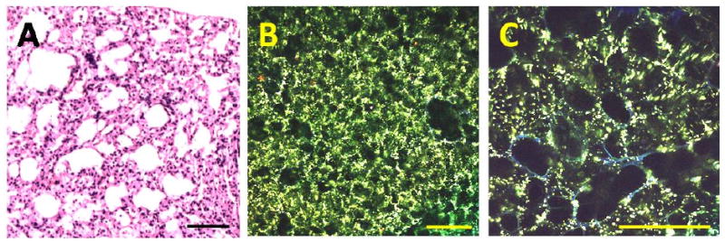
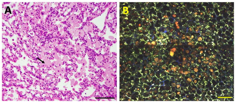
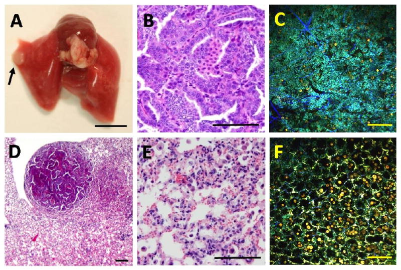
Lung cancer is the leading killer among all cancers for both men and women in the US, and is associated with one of the lowest 5-year survival rates. Current diagnostic techniques, such as histopathological assessment of tissue obtained by computed tomography guided biopsies, have limited accuracy, especially for small lesions. Early diagnosis of lung cancer can be improved by introducing a real-time, optical guidance method based on the in vivo application of multiphoton microscopy (MPM). In particular, we hypothesize that MPM imaging of living lung tissue based on two-photon excited intrinsic fluorescence and second harmonic generation can provide sufficient morphologic and spectroscopic information to distinguish between normal and diseased lung tissue. Here, we used an experimental approach based on MPM with multichannel fluorescence detection for initial discovery that MPM spectral imaging could differentiate between normal and neoplastic lung in ex vivo samples from a murine model of lung cancer. Current results indicate that MPM imaging can directly distinguish normal and neoplastic lung tissues based on their distinct morphologies and fluorescence emission properties in non-processed lung tissue. Moreover, we found initial indication that MPM imaging differentiates between normal alveolar tissue, inflammatory foci, and lung neoplasms. Our long-term goal is to apply results from ex vivo lung specimens to aid in the development of multiphoton endoscopy for in vivo imaging of lung abnormalities in various animal models, and ultimately for the diagnosis of human lung cancer.
Figures
References
-
- Jemal A, Siegel R, Ward E, Hao YP, Xu JQ, Murray T, Thun MJ. Cancer statistics, 2008. Ca-Cancer J Clin. 2008;58:71–96. - PubMed
-
- Schreiber G, McCrory DC. Performance characteristics of different modalities for diagnosis of suspected lung cancer - Summary of published evidence. Chest. 2003;123:115S–128S. - PubMed
-
- Montaudon M, Latrabe V, Pariente A, Corneloup O, Begueret H, Laurent F. Factors influencing accuracy of CT-guided percutaneous biopsies of pulmonary lesions. Eur Radiol. 2004;14:1234–1240. - PubMed
-
- Meyer C. Transthoracic Needle Aspiration Biopsy of Benign and Malignant Lung Lesions—A Commentary. AJR. 2007;188:891–893. - PubMed
-
- Khouri NF, Stitik FP, Erozan YS, Gupta PK, Kim WS, Scott WW, Hamper UM, Mann RB, Eggleston JC, Baker RR. Transthoracic needle aspiration biopsy of benign and malignant lung lesions. AJR. 1985;144:281–288. - PubMed
Grants and funding
LinkOut - more resources
Full Text Sources