

White matter in aging and cognition: a cross-sectional study of microstructure in adults aged eighteen to eighty-three
- PMID: 20446132
- PMCID: PMC2895988
- DOI: 10.1080/87565641003696775
White matter in aging and cognition: a cross-sectional study of microstructure in adults aged eighteen to eighty-three
Abstract
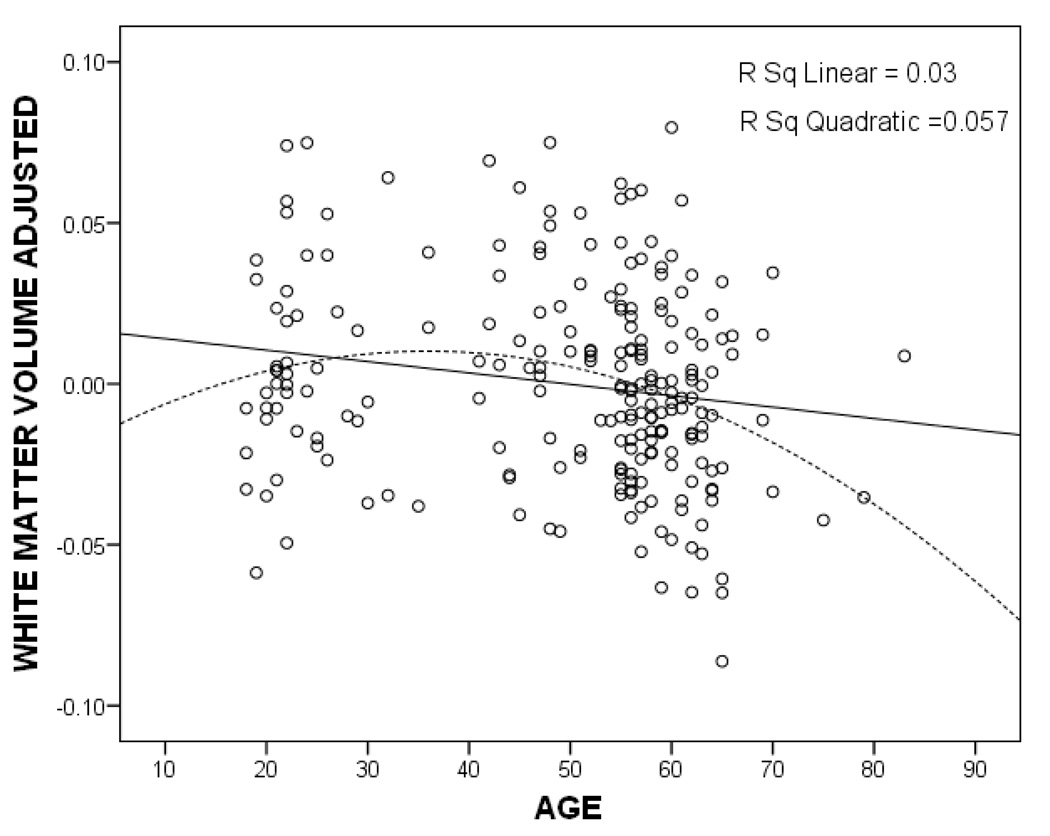
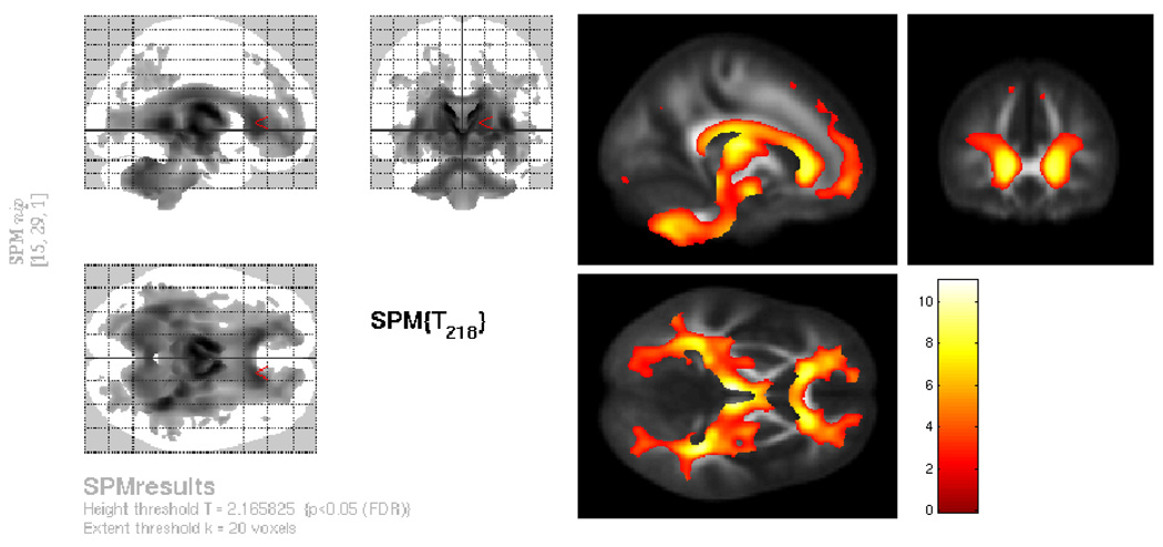
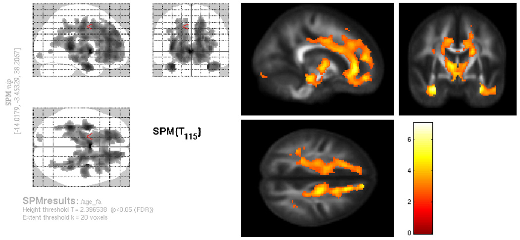
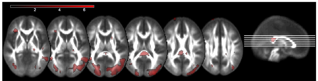
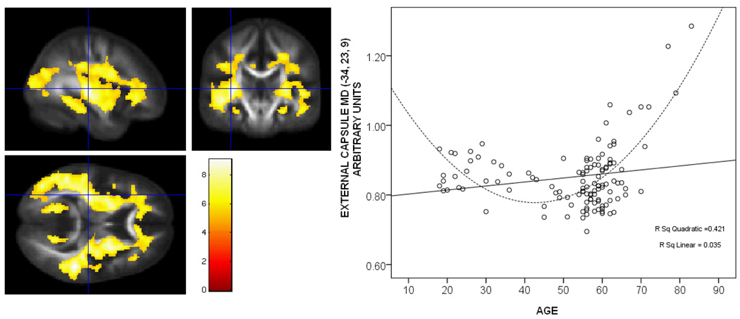
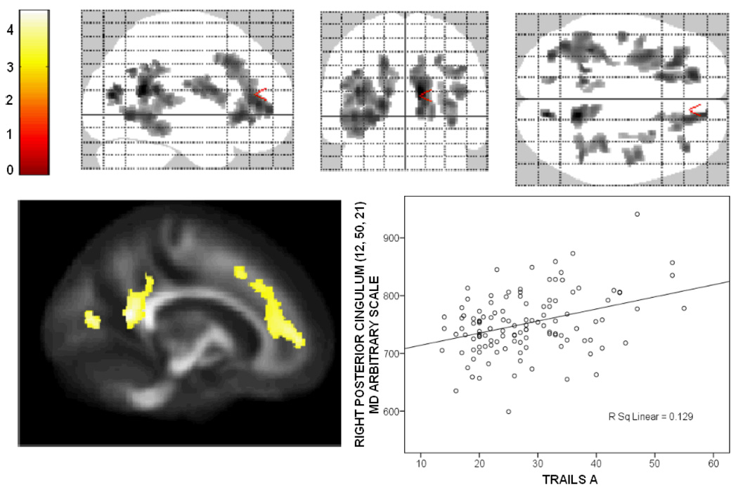
Structural brain change and concomitant cognitive decline are the seemingly unavoidable escorts of aging. Despite accumulating studies detailing the effects of age on the brain and cognition, the relationship between white matter features and cognitive function in aging have only recently received attention and remain incompletely understood. White matter microstructure can be measured with diffusion tensor imaging (DTI), but whether DTI can provide unique information on brain aging that is not explained by white matter volume is not known. In the current study, the relationship between white matter microstructure, age, and neuropsychological function was assessed using DTI in a statistical framework that employed white matter volume as a voxel-wise covariate in a sample of 120 healthy adults across a broad age range (18-83). Memory function and executive function were modestly correlated with the DTI measures while processing speed showed the greatest extent of correlation. The results suggest that age-related white matter alterations underlie age-related declines in cognitive function. Mean diffusivity and fractional anisotropy in several white matter brain regions exhibited a nonlinear relationship with age, while white matter volume showed a primarily linear relationship with age. The complex relationships between cognition, white matter microstructure, and white matter volume still require further investigation.
Figures
References
-
- Abe O, Aoki S, Hayashi N, Yamada H, Kunimatsu A, Mori H, et al. Normal aging in the central nervous system: quantitative MR diffusion-tensor analysis. Neurobiology of Aging. 2002;23(3):433–441. - PubMed
-
- Aggleton JP, Hunt PR, Nagle S, Neave N. The effects of selective lesions within the anterior thalamic nuclei on spatial memory in the rat. Behavioural Brain Research. 1996;81(1–2):189–198. - PubMed
-
- Allen JS, Damasio H, Grabowski TJ, Bruss J, Zhang W. Sexual dimorphism and asymmetries in the gray-white composition of the human cerebrum. Neuroimage. 2003;18(4):880–894. - PubMed
-
- Au R, Massaro JM, Wolf PA, Young ME, Beiser A, Seshadri S, et al. Association of white matter hyperintensity volume with decreased cognitive functioning: the Framingham Heart Study. Archives of Neurology. 2006;63(2):246–250. - PubMed
-
- Bartzokis G, Beckson M, Lu PH, Nuechterlein KH, Edwards N, Mintz J. Age-related changes in frontal and temporal lobe volumes in men: a magnetic resonance imaging study. Archives of General Psychiatry. 2001;58(5):461–465. - PubMed
Publication types
MeSH terms
Grants and funding
LinkOut - more resources
Full Text Sources
Medical