

Diffusion tensor imaging of the cingulum bundle in children after traumatic brain injury
- PMID: 20446136
- PMCID: PMC3229222
- DOI: 10.1080/87565641003696940
Diffusion tensor imaging of the cingulum bundle in children after traumatic brain injury
Abstract
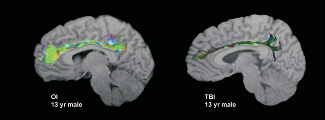
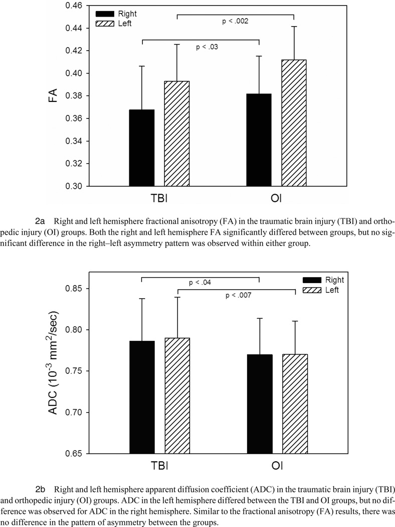
Structural damage to the prefrontal-cingulate network has been implicated in cognitive and neurobehavioral deficits associated with traumatic brain injury (TBI). Forty-six children who had sustained moderate-to-severe TBI and 43 children with extracranial injury were imaged using diffusion tensor imaging (DTI). Decreased fractional anisotropy (FA) and increased apparent diffusion coefficient (ADC) values were found in the cingulum bundles bilaterally in the TBI group. Cingulum ADC was related to frontal lesion volume, injury severity, and injury mechanism. Finally, cingulum DTI parameters were related to cognitive control measures. DTI detects TBI-related injury to the cingulum, which may facilitate advances in assessment and treatment.
Figures
References
-
- Ashwal S, Holshouser BA, Tong KA. Use of advanced neuroimaging techniques in the evaluation of pediatric traumatic brain injury. Developmental Neuroscience. 2006;28(4–5):309–326. - PubMed
-
- Azouvi P. Neuroimaging correlates of cognitive and functional outcome after traumatic brain injury. Current Opinion of Neurology. 2000;13(6):665–669. - PubMed
-
- Benson RR, Meda SA, Vasudevan S, Kou Z, Govindarajan KA, Hanks RA, et al. Global white matter analysis of diffusion tensor images is predictive of injury severity in traumatic brain injury. Journal of Neurotrauma. 2007;24(3):446–459. - PubMed
Publication types
MeSH terms
Grants and funding
LinkOut - more resources
Full Text Sources