

Nanoliter multiplex PCR arrays on a SlipChip
- PMID: 20446698
- PMCID: PMC2916686
- DOI: 10.1021/ac1007249
Nanoliter multiplex PCR arrays on a SlipChip
Abstract
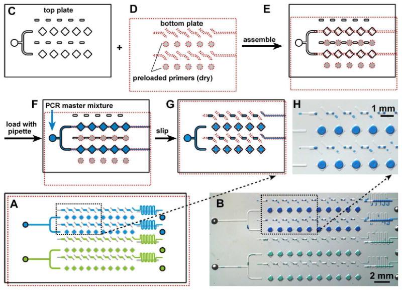
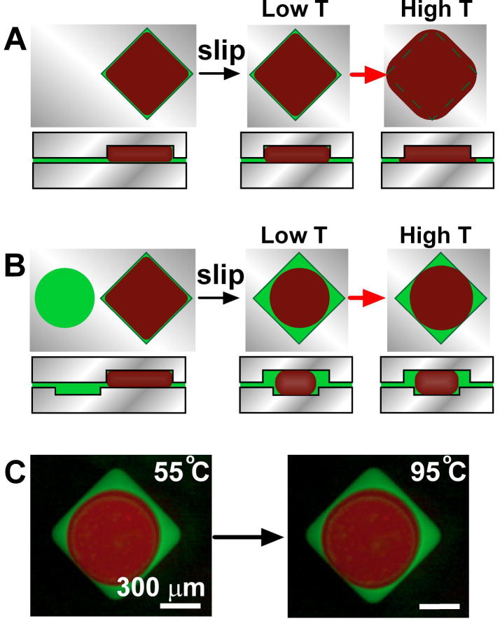
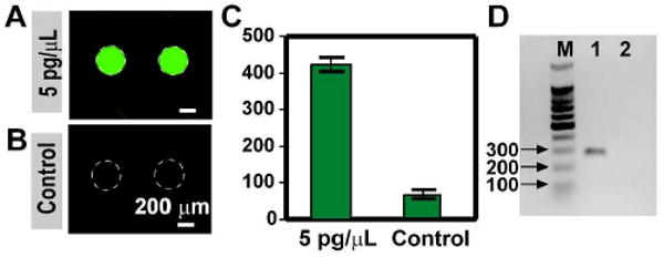
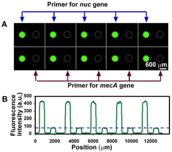
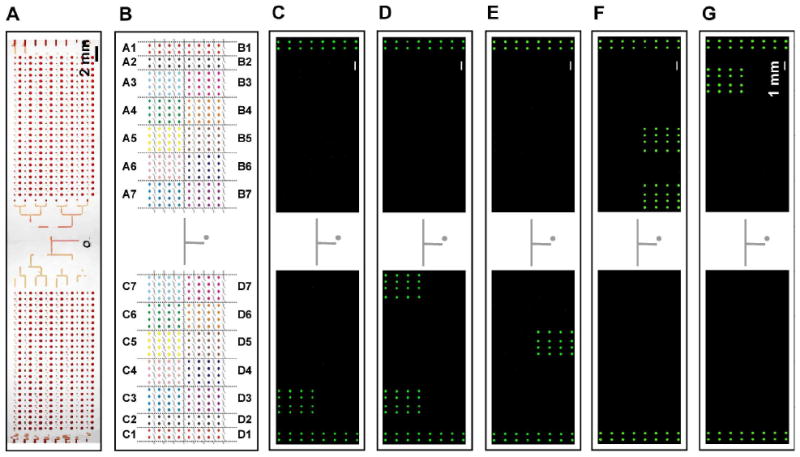
The SlipChip platform was tested to perform high-throughput nanoliter multiplex PCR. The advantages of using the SlipChip platform for multiplex PCR include the ability to preload arrays of dry primers, instrument-free sample manipulation, small sample volume, and high-throughput capacity. The SlipChip was designed to preload one primer pair per reaction compartment and to screen up to 384 different primer pairs with less than 30 nanoliters of sample per reaction compartment. Both a 40-well and a 384-well design of the SlipChip were tested for multiplex PCR. In the geometries used here, the sample fluid was spontaneously compartmentalized into discrete volumes even before slipping of the two plates of the SlipChip, but slipping introduced additional capabilities that made devices more robust and versatile. The wells of this SlipChip were designed to overcome potential problems associated with thermal expansion. By using circular wells filled with oil and overlapping them with square wells filled with the aqueous PCR mixture, a droplet of aqueous PCR mixture was always surrounded by the lubricating fluid. In this design, during heating and thermal expansion, only oil was expelled from the compartment and leaking of the aqueous solution was prevented. Both 40-well and 384-well devices were found to be free from cross-contamination, and end point fluorescence detection provided reliable readout. Multiple samples could also be screened on the same SlipChip simultaneously. Multiplex PCR was validated on the 384-well SlipChip with 20 different primer pairs to identify 16 bacterial and fungal species commonly presented in blood infections. The SlipChip correctly identified five different bacterial or fungal species in separate experiments. In addition, the presence of the resistance gene mecA in methicillin resistant Staphylococcus aureus (MRSA) was identified. The SlipChip will be useful for applications involving PCR arrays and lays the foundation for new strategies for diagnostics, point-of-care devices, and immobilization-based arrays.
Figures
Similar articles
-
Digital isothermal quantification of nucleic acids via simultaneous chemical initiation of recombinase polymerase amplification reactions on SlipChip.Anal Chem. 2011 May 1;83(9):3533-40. doi: 10.1021/ac200247e. Epub 2011 Apr 8. Anal Chem. 2011. PMID: 21476587 Free PMC article.
-
Digital PCR on a SlipChip.Lab Chip. 2010 Oct 21;10(20):2666-72. doi: 10.1039/c004521g. Epub 2010 Jul 1. Lab Chip. 2010. PMID: 20596567 Free PMC article.
-
Multiplex Digital Polymerase Chain Reaction on a Droplet Array SlipChip for Analysis of KRAS Mutations in Pancreatic Cancer.ACS Sens. 2023 Jan 27;8(1):114-121. doi: 10.1021/acssensors.2c01776. Epub 2022 Dec 15. ACS Sens. 2023. PMID: 36520653
-
SlipChip for immunoassays in nanoliter volumes.Anal Chem. 2010 Apr 15;82(8):3276-82. doi: 10.1021/ac100044c. Anal Chem. 2010. PMID: 20334360 Free PMC article.
-
Folic acid supplementation and malaria susceptibility and severity among people taking antifolate antimalarial drugs in endemic areas.Cochrane Database Syst Rev. 2022 Feb 1;2(2022):CD014217. doi: 10.1002/14651858.CD014217. Cochrane Database Syst Rev. 2022. PMID: 36321557 Free PMC article.
Cited by
-
Polymerase Chain Reaction Chips for Biomarker Discovery and Validation in Drug Development.Micromachines (Basel). 2025 Feb 20;16(3):243. doi: 10.3390/mi16030243. Micromachines (Basel). 2025. PMID: 40141854 Free PMC article. Review.
-
Digital isothermal quantification of nucleic acids via simultaneous chemical initiation of recombinase polymerase amplification reactions on SlipChip.Anal Chem. 2011 May 1;83(9):3533-40. doi: 10.1021/ac200247e. Epub 2011 Apr 8. Anal Chem. 2011. PMID: 21476587 Free PMC article.
-
Evaluation of a high-throughput, microfluidics platform for performing TaqMan™ qPCR using formalin-fixed paraffin-embedded tumors.Bioanalysis. 2013 Jul;5(13):1623-33. doi: 10.4155/bio.13.125. Bioanalysis. 2013. PMID: 23822126 Free PMC article.
-
Microfluidic compartmentalization to identify gene biomarkers of infection.Biomicrofluidics. 2020 Dec 3;14(6):061502. doi: 10.1063/5.0032849. eCollection 2020 Nov. Biomicrofluidics. 2020. PMID: 33312326 Free PMC article. Review.
-
Advancing microfluidic diagnostic chips into clinical use: a review of current challenges and opportunities.Lab Chip. 2022 Aug 23;22(17):3110-3121. doi: 10.1039/d2lc00024e. Lab Chip. 2022. PMID: 35674283 Free PMC article. Review.
References
-
- Kambouris M, Jackson CE, Feldman GL. Hum Mutat. 1996;8:64–70. - PubMed
-
- Casilli F, Di Rocco ZC, Gad S, Tournier I, Stoppa-Lyonnet D, Frebourg T, Tosi M. Hum Mutat. 2002;20:218–226. - PubMed
-
- Westh H, Lisby G, Breysse F, Boddinghaus B, Chomarat M, Gant V, Goglio A, Raglio A, Schuster H, Stuber F, Wissing H, Hoeft A. Clin Microbiol Infect. 2009;15:544–551. - PubMed
Publication types
MeSH terms
Grants and funding
LinkOut - more resources
Full Text Sources
Other Literature Sources
