Review
. 2010 Oct;13(8):1273-96.
doi: 10.1089/ars.2010.3109.

Conformational plasticity and structure/function relationships in cytochromes P450

Affiliations
Review

Conformational plasticity and structure/function relationships in cytochromes P450

Thomas C Pochapsky et al. Antioxid Redox Signal. 2010 Oct.

Abstract

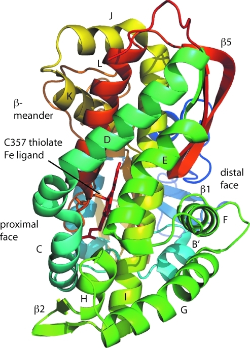
The cytochrome P450s are a superfamily of enzymes that are found in all kingdoms of living organisms, and typically catalyze the oxidative addition of atomic oxygen to an unactivated C-C or C-H bond. Over 8000 nonredundant sequences of putative and confirmed P450 enzymes have been identified, but three-dimensional structures have been determined for only a small fraction of these. While all P450 enzymes for which structures have been determined share a common global fold, the flexibility and modularity of structure around the active site account for the ability of P450 enzymes to accommodate a vast number of structurally dissimilar substrates and support a wide range of selective oxidations. In this review, known P450 structures are compared, and some structural criteria for prediction of substrate selectivity and reaction type are suggested. The importance of dynamic processes such as redox-dependent and effector-induced conformational changes in determining catalytic competence and regio- and stereoselectivity is discussed, and noncrystallographic methods for characterizing P450 structures and dynamics, in particular, mass spectrometry and nuclear magnetic resonance spectroscopy are reviewed.


Figures

FIG. 1.
FIG. 1.
Structure of camphor hydroxylase CYP101 (cytochrome P450cam) from PDB entry 2CPP, as viewed from the distal face. Secondary structural features are labeled according to the scheme of Poulos (109). Secondary structural features are color-coded from N-terminal (blue) to C-terminal (red). The directional arrow marked “N” indicates the “north” described in the text. (For interpretation of the references to color in this figure legend, the reader is referred to the web version of this article at www.liebertonline.com/ars). Except where noted, all figures were generated using PyMOL (20). (For interpretation of the references to color in this figure legend, the reader is referred to the web version of this article at www.liebertonline.com/ars).
FIG. 2.
FIG. 2.
Structure of CYP101 (cytochrome P450cam) from PDB entry 2CPP, as viewed from the “north” face (top of structure in Fig. 1). Distal and proximal features refer to position with respect to the plane of the heme. Secondary structural features are color-coded from N-terminal (blue) to C-terminal (red). (For interpretation of the references to color in this figure legend, the reader is referred to the web version of this article at www.liebertonline.com/ars).
FIG. 3.
FIG. 3.
Structure of fatty acid ω-2 hydroxylase CYP102 (cytochrome P450BM3) from PDB entry 1BVY (133), as viewed from the distal face. Secondary structural features are labeled in analogy to structure of CYP101 (Fig. 1). Secondary structural features are color-coded from N-terminal (blue) to C-terminal (red). Approximate position of the substrate binding channel as reported by Ravichandran et al. is indicated by a dotted line (119). (For interpretation of the references to color in this figure legend, the reader is referred to the web version of this article at www.liebertonline.com/ars).
FIG. 4.
FIG. 4.
CYP108 (cytochrome P450terp), from PDB entry 1CPT (37), viewed from distal face in the same orientation as Figures 1 and 3 (see directional arrow). Note the absence of the F–G loop, which is disordered in the crystal. (For interpretation of the references to color in this figure legend, the reader is referred to the web version of this article at www.liebertonline.com/ars).
FIG. 5.
FIG. 5.
Cytochrome P450 EpoK (PDB entry 1Q5D, (87)) viewed from distal face in the same orientation as CYP101 in Figure 1. Substrate (epothilone B, labeled Epo) (For interpretation of the references to color in this figure legend, the reader is referred to the web version of this article at www.liebertonline.com/ars).
FIG. 6.
FIG. 6.
Active site of P450 OxyB (PDB entry 1LFK, (167)) viewed from distal face in the same orientation as CYP101 in Figure 1. Unusually, most of the expected primary contacts for substrate vancomycin are prolines. (For interpretation of the references to color in this figure legend, the reader is referred to the web version of this article at www.liebertonline.com/ars).
FIG. 7.
FIG. 7.
Substrate-free CYP2C5 structure (1DT6 (47)) viewed from the distal face in the same orientation as CYP101 in Figure 1. Note that the F–G loop is disordered and there is no regular secondary structure in the B–C loop. The approximate location of the membrane binding interface including an N-terminal helix (not present) and portions of the F–G loop is marked as MBI. (For interpretation of the references to color in this figure legend, the reader is referred to the web version of this article at www.liebertonline.com/ars).
FIG. 8.
FIG. 8.
CYP2C5 with substrate dimethylsulfathiazole (DMZ) bound in two orientations (1N6B (47)). The active site is viewed from the south edge. Note that the F–G loop is ordered and a short B' helix is present, unlike the substrate-free form (Fig. 7). (For interpretation of the references to color in this figure legend, the reader is referred to the web version of this article at www.liebertonline.com/ars).
FIG. 9.
FIG. 9.
Structure of CYP3A4 determined with inhibitor ketoconazole bound (2V0M (25)) viewed from the distal face in the same orientation as CYP101 in Figure 1. Note that the extended F–G loop with two helical regions F' and G'. (For interpretation of the references to color in this figure legend, the reader is referred to the web version of this article at www.liebertonline.com/ars).
FIG. 10.
FIG. 10.
Active site of CYP1A2 with substrate naphthoflavone bound (2HI4, (126)). Naphthoflavone carbon atoms are shown in purple. (For interpretation of the references to color in this figure legend, the reader is referred to the web version of this article at www.liebertonline.com/ars).
FIG. 11.
FIG. 11.
Comparison of the cis (left) and trans (right) conformations of the Ile 88-Pro 89 amide bond initiating the B' helix in CYP101. Structures shown are based on molecular dynamics simulations supported by NMR experiments (5). Pro 89 is shown in orange, Ile 395 in light blue, Thr 185 in dark blue, and camphor (substrate) is in magenta. Note the greater solvent exposure of camphor in the trans conformation, indicating a more accessible active site. (For interpretation of the references to color in this figure legend, the reader is referred to the web version of this article at www.liebertonline.com/ars).
FIG. 12.
FIG. 12.
Active site structure of prostacyclin synthase, CYP8A1 (PDB ID 3B99, (68)) with substrate analog 9,11-azoprosta-5(Z),13(E)-dien-1-oic acid bound (labeled U51 in figure). (For interpretation of the references to color in this figure legend, the reader is referred to the web version of this article at www.liebertonline.com/ars).
FIG. 13.
FIG. 13.
Anchoring of β3 strand at the south edge of the active site by a salt bridge between the heme propionate and conserved Arg residues in (a) CYP101 (2CPP, (109)), and (b) P450 epoK (1Q5D, (87)). Substrate contact residues in register with the conserved Arg are Val 295 in CYP101 and Thr 305 in P450 EpoK. Substrates are camphor in CYP101 and epothilone B (epoB) in EpoK. See text for complete discussion. In (c) CYP2C5 (1NR6, (156)), the Arg is not present in the β3 strand, but the salt bridge is conserved via Arg 430 from the proximal side of the enzyme. (For interpretation of the references to color in this figure legend, the reader is referred to the web version of this article at www.liebertonline.com/ars).
FIG. 14.
FIG. 14.
Active sites of two P450 enzymes that bind cholesterol-derived substrates in opposite orientations. (Left) Active site of CYP46A1 with cholesterol sulfate bound (2Q9F, (79)). Side chains in dark blue are from the A helix, light blue from the B–C loop, green from the F helix and F–G loop, orange from the β3 strand and loop, and red from the β5 loop. (Right) Active site of aromatase with substrate androstenedione bound (CYP19A1, PDB ID 3EQM, (29)). Side chains in light blue from the B–C loop, green from the F helix, orange from the β3 strand and loop, and red from the β5 loop. Note the unusual Pro in the I helix, P308, which appears to make the “kink” often found in the I helix providing a binding site for bound O2. (For interpretation of the references to color in this figure legend, the reader is referred to the web version of this article at www.liebertonline.com/ars).
FIG. 15.
FIG. 15.
View from west side of the active site of CYP107L1 (PikC) from structure 2WHW (67) showing side chains from the I helix, β3 and β5 loops involved in binding a substrate modified with a desaminoglucoside (deso) to convert a C13 macrocycle into a substrate for oxidation by this enzyme. The desaminoglucoside binding site is defined by Tyr 295 and Thr 294 on the β3 loop and Met 394 on the β5 loop. The desaminoglucoside is also contacted by Glu 94 on the B–C loop and Phe 178 on the F helix (not shown). Other marked residues (Ile 239 and Val 242 on the I helix, and Val 290 on the east wall of the active site) contact the macrocycle. (For interpretation of the references to color in this figure legend, the reader is referred to the web version of this article at www.liebertonline.com/ars).
FIG. 16.
FIG. 16.
Alignment of β3 regions lining the active sites of P450 enzymes discussed in the text. Numbers to the right of the aligned sequences are sequence number of the aligned Arg/His residues from each sequence.
FIG. 17.
FIG. 17.
Active site of CYP119, from S. solfataricus (PDB ID I1O8, (97)), showing positions of active site residues proposed to be important for substrate recognition and binding. Translucent oval shows approximate position proposed for bound substrate. (For interpretation of the references to color in this figure legend, the reader is referred to the web version of this article at www.liebertonline.com/ars).
FIG. 18.
FIG. 18.
Partial sequence alignments of selected putative P450 sequences with cytochrome P450cam (CYP101) from P. putida showing correlations discussed in text. Secondary structural features are noted above the CAM sequence. GenBank accession numbers are as follows: ZP_01304513.1 Sphingomonas sp. SKA58, YP_495795.1 Novosphingobium aromaticivorans, ZP_04956740.1 gamma proteobacterium, YP_001774457.1 Burkholderia cenocepacia, YP_001262244.1 Sphingomonas wittichii. Alignments were made using the BLAST alignment tool (blast.ncbi.nlm.nih.gov) (2). (For interpretation of the references to color in this figure legend, the reader is referred to the web version of this article at www.liebertonline.com/ars).

Similar articles

Cited by

References

    1. Aldag C. Gromov IA. Garcia–Rubio I. von Koenig K. Schlichting I. Jaun B. Hilvert D. Probing the role of the proximal heme ligand in cytochrome P450cam by recombinant incorporation of selenocysteine. Proc Natl Acad Sci USA. 2009;106:5481–5486. - PMC - PubMed
    1. .Altschul SF.Madden TL.Schaffer AA.Zhang JH.Zhang Z.Miller W.Lipman DJ.Gapped BLAST and PSI-BLAST: A new generation of protein database search programs Nucleic Acids Res 253389–3402.1997. - PMC - PubMed
    1. Andersen JF. Tatsuta K. Gunji H. Ishiyama T. Hutchinson CR. Substrate-apecificity of 6-Deoxyerythronolide-B hydroxylase, a bacterial cytochrome P450 of erythromycin-a biosynthesis. Biochemistry. 1993;32:1905–1913. - PubMed
    1. Arimoto R. Computational models for predicting interactions with cytochrome P450 enzyme. Current Topics Med Chem. 2006;6:1609–1618. - PubMed
    1. Asciutto EK. Madura JD. Pochapsky SS. OuYang B. Pochapsky TC. Structural and dynamic implications of an effector-induced backbone amide cis-trans isomerization in cytochrome P450(cam) J Mol Biol. 2009;388:801–814. - PMC - PubMed

Publication types

MeSH terms

Substances

LinkOut - more resources