Review
. 2010 Jun;45(3):215-32.
doi: 10.3109/10409231003796523.

The tertiary structure of group II introns: implications for biological function and evolution

Affiliations
Review

The tertiary structure of group II introns: implications for biological function and evolution

Anna Marie Pyle. Crit Rev Biochem Mol Biol. 2010 Jun.

Abstract

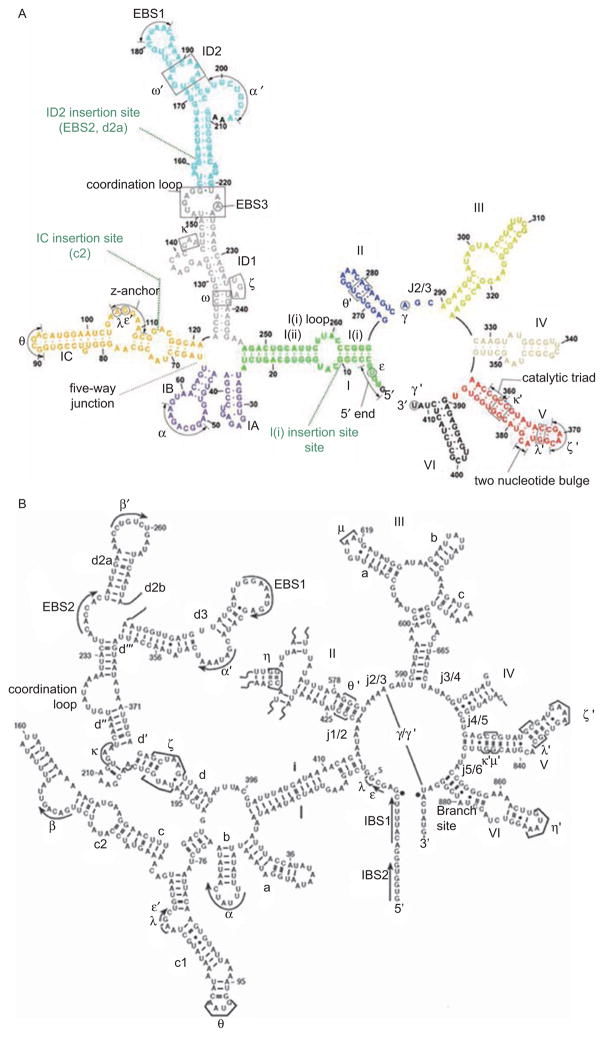
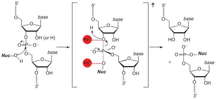
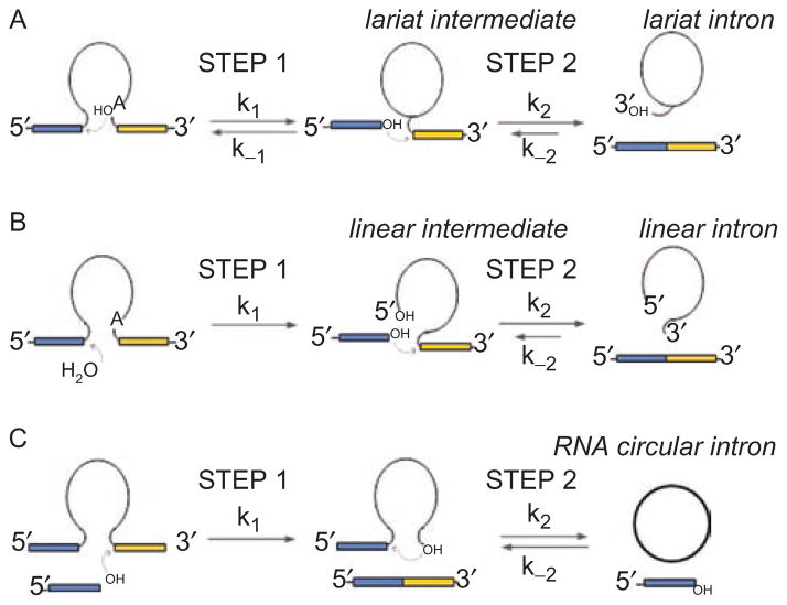
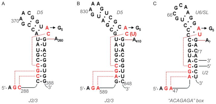
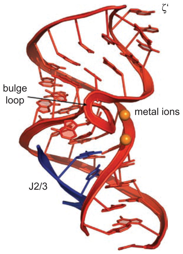
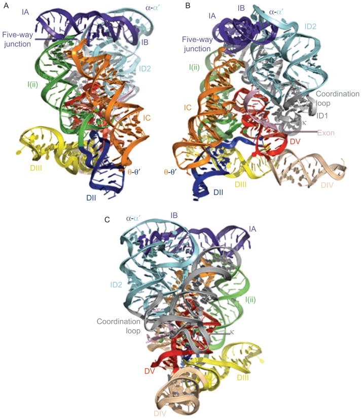
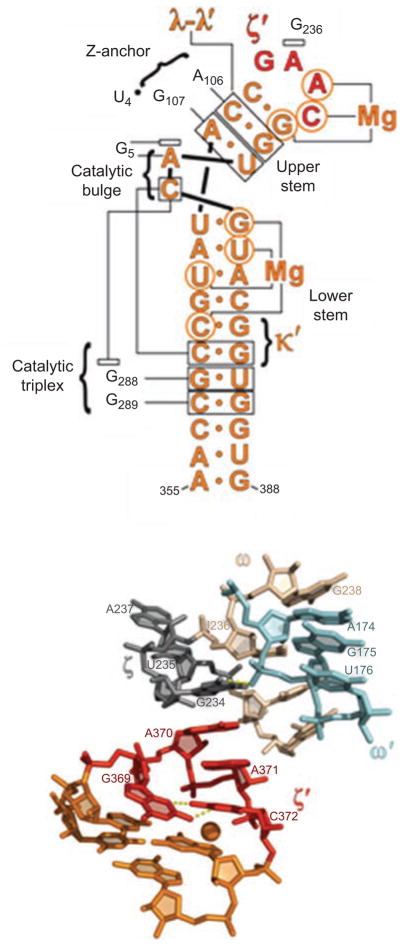
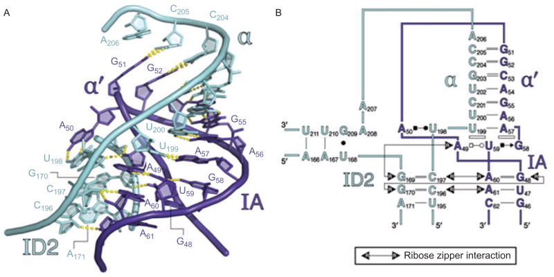
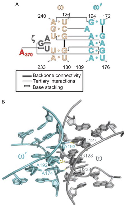
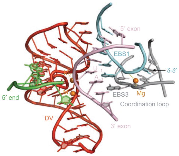
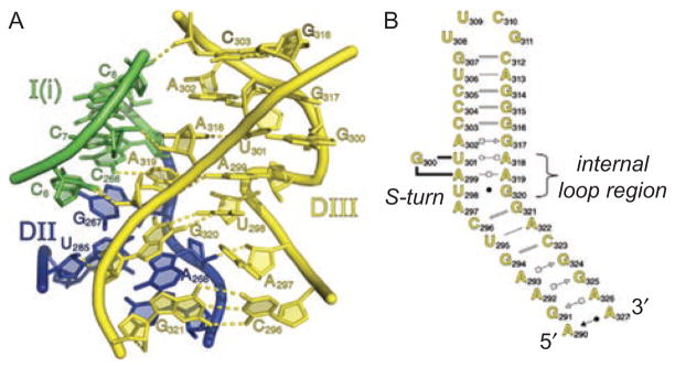
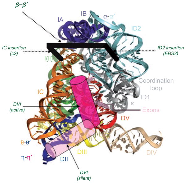
Group II introns are some of the largest ribozymes in nature, and they are a major source of information about RNA assembly and tertiary structural organization. These introns are of biological significance because they are self-splicing mobile elements that have migrated into diverse genomes and played a major role in the genomic organization and metabolism of most life forms. The tertiary structure of group II introns has been the subject of many phylogenetic, genetic, biochemical and biophysical investigations, all of which are consistent with the recent crystal structure of an intact group IIC intron from the alkaliphilic eubacterium Oceanobacillus iheyensis. The crystal structure reveals that catalytic intron domain V is enfolded within the other intronic domains through an elaborate network of diverse tertiary interactions. Within the folded core, DV adopts an activated conformation that readily binds catalytic metal ions and positions them in a manner appropriate for reaction with nucleic acid targets. The tertiary structure of the group II intron reveals new information on motifs for RNA architectural organization, mechanisms of group II intron catalysis, and the evolutionary relationships among RNA processing systems. Guided by the structure and the wealth of previous genetic and biochemical work, it is now possible to deduce the probable location of DVI and the site of additional domains that contribute to the function of the highly derived group IIB and IIA introns.


Conflict of interest statement

Declaration of interest

I am an Investigator with the Howard Hughes Medical Institute, which supported this work. This work was also supported by a generous grant from the National Institutes of Health (GM50313). The author report no conflicts of interest. The author alone is responsible for the content and writing of the paper.

Figures

Figure 1
Figure 1
Secondary structures of smaller (Group IIC) and larger (Group IIB) intron variants. (A) Secondary structure of the crystallized group IIC intron construct from Oceanobacillus iheyensis (O.i.). The sequence of the full-length O.i. intron is provided in Figure S2 of Toor et al. (2008). Domains are indicated with roman numerals and long-range interactions are indicated by Greek letters. Color coding is the same as shown in Figure 6, for the three-dimensional structure. Probable sites of the ID2, IC and I(i) insertions are indicated with green dotted lines, as indicated. (B) Secondary structure of the ai5γ Group IIB intron from the mitochondrial genome of Saccharomyces cerevisiae. Domains are indicated with roman numerals and subdomains with lower case letters. Long-range tertiary interaction partners are indicated with Greek letters. Note that EBS1 and EBS2 form Watson–Crick base pairs with IBS1 and IBS2 of the 5′-exon, respectively. Exon/intron boundaries are indicated by black dots.
Figure 2
Figure 2
The chemical mechanism of catalysis by group II introns. This is the basic reaction underlying both steps of splicing and the reverse-splicing reactions involved in intron mobility. In this scheme, “Nuc” can represent the 2′-OH group of the branch-site, the 3′-OH group of the 5′-exon or the terminus of the intron, and it can also represent water. Note that the 2′-group on the departing sugar moiety can be a 2′-hydroxyl group (for RNA) or a 2′-hydrogen (for DNA), as the intron can react with either type of substrate. Metal ions are indicated with red balls, and they are in the positions indicated through biochemical work, and implicated by the position of metal ions in the crystal structure, as shown in Figure 11.
Figure 3
Figure 3
Three pathways for group II intron splicing and intron excision. (A) The branching pathway that leads to lariat formation. (B) The hydrolytic pathway that leads to release of linear intron. Note that the reverse reactions shown for (A) and (B) are the basis for the intron mobility reaction, whereby group II introns insert themselves into the sense-strand of target DNA (Pyle and Lambowitz, 2006). (C) A possible circle formation pathway. This reaction may be initiated by a free 5′-exon that has been liberated through the spliced-exon reopening reaction on a different intron boundary. Note that 5′-exons are shown in blue and 3′-exons are shown in yellow. The intron is indicated by a thin black line.
Figure 4
Figure 4
Secondary structures of DV and the U6 snRNA, together with anticipated contacts within the active-site. (A) The DV and J2/3 sequences from group IIC intron Oceanobacillus iheyensis, with active-site tertiary interactions determined from the crystal structure (Keating et al., 2010). The J2/3 nucleotides form a major groove triplex with the “catalytic CGC triad” of conserved nucleotides at the base of DV, and with C259 from the bulge. (B) The DV and J2/3 sequences from group IIB intron ai5γ. Anticipated long-range interactions are shown, by analogy to the O.i. crystal structure. These can be readily modeled into the active-site of the solved structure (Keating et al., 2010). (C) The U6 snRNA from the human spliceosome. The ACAGAGA box is hypothesized to function like J2/3, forming a triple helix that supports the metal-binding, actives-site bulge at positions 73 and 74. The U6 bulge region is hypothesized to form tertiary interactions similar to those observed within the IIC intron (Keating et al., 2010). Reprinted with permission from RNA (Keating et al., 2010).
Figure 5
Figure 5
The structure of DV within the catalytic core of the folded group IIC intron. DV nucleotides are shown in red. Two metal ions (orange spheres) are bound near the tightly twisted bulge loop. The J2/3 nucleotides (blue ribbon) form a triple helix with the major groove at the base of DV. The site for ζ–ζ′ interaction with DI is indicated. This image was rendered from pdb code 3IGI.
Figure 6
Figure 6
Three views of the molecular structure of the Oceanobacillus iheyensis group IIC intron (Toor et al., 2010), color-coded and labeled as shown in Figure 1A. This image was rendered from pdb code 3IGI. Reprinted with permission from RN (Toor et al., 2010).
Figure 7
Figure 7
The network of interactions involving DV. (A) A secondary structural map of the interactions observed between DV and the rest of the intron. Brackets indicate sites of long-range interaction and contacts with metals, open rectangles indicate stacked nucleobases, labels indicate identity of tertiary interactions. Red nucleotides are involved in the ζ–ζ′ interaction. (B) The molecular structure of the ζ–ζ′ interaction, determined from the crystal structure of the Oceanobacillus iheyensis group IIC intron. Unlike most tetraloop receptors, this interaction is dictated only by a single stack between A370 of DV and G236 in DI. The unusual tetraloop contains a bound metal ion. The receptor is buttressed by the ω–ω′ interaction that is idiosynchratic to IIC introns (Keating et al., 2008). First published in J Mol Biol (2008) 283, 475–481; Figures 1a and 2a, pages 476 and 478 respectively.
Figure 8
Figure 8
Generalized tertiary folding pathway of the group IIB intron ai5γ. After forming the basic secondary structure (defined here as U), domain I (blue cylinders) folds first, forming an intermediate (Ic) in which the catalytic domains are not docked within the core. The Ic–N transition involves the docking of D5 and D3. First published in TIBS (2007) 32, 138–145; Figure 3a, page 142.
Figure 9
Figure 9
The α–α′ interaction and its supporting ribose zipper network. (A) Molecular structure of α–α′, showing that the strands beneath it are joined through sequential contacts between 2′-hydroxyl groups (ribose zippers). (B) Secondary structural representation of the same region. Open rectangles indicate base stacking interactions. Types of base-pairing are represented using the Leontis–Westhof symbolism. Color coding is as shown in Figure 1A and 6. Reprinted with permission from RNA (Toor et al., 2010).
Figure 10
Figure 10
The ω–ω′ interaction of group IIC introns. (A) Secondary structural representation of this sequence-specific ribose zipper motif. Color coding is as shown in Figures 1A and 6. First published in J Mol Biol (2008) 283, 475–481; Figure 1b, page 476. (B) Molecular structure of the ω–ω′ region within the Oceanobacillus iheyensis group IIC intron, showing the ribose zipper hydrogen bonds in yellow. First published in Science (2008) 320, 77–82; Figure 3c, page 79.
Figure 11
Figure 11
Exon recognition surface within the Oceanobacillus iheyensis group IIC intron. Bound target oligonucleotide (pink) interacts with the intron core through base-pairing interactions with EBS1 (blue) and EBS3 (gray), which are joined together and form one structural unit that is stabilized through the δ–δ′ interaction and a bridging metal ion. Note that the result is a duplex in which EBS3 stacks beneath EBS1, as they both base pair simultaneously with the oligonucleotide. Rendered from pdb code 3IGI.
Figure 12
Figure 12
The internal loop in DIII dictates placement of the DI and DII stems. (A) The molecular structure of the DIII loop region (yellow), showing that it forms extensive interactions with the bottom of the DI (blue) and DII (green) stems, helping the junction of these coaxially stacked stems to become properly oriented within the structure. Rendered from pdb code 3IGI. (B) Secondary structure of DIII from Oceanobacillus iheyensis, as determined from the crystal structure. Color coding is the same as in Figures 1A and 6. Reprinted with permission from RNA (Toor et al., 2010).
Figure 13
Figure 13
The hypothetical position of DVI, EBS2 and β–β′. Superimposed on a model of the group II intron crystal structure (rendered from pdb file 3IGI), the proposed position of various derived domains is shown. DVI is likely to have two functional conformations. The “active” conformation that participates in branching is likely to be in the position indicated by the dark pink cylinder. The “silent” conformation is stabilized by the η–η′ interaction between DVI and DII is likely to be located in the position indicated by the light pink cylinder. DVI probably transits between these two sites in order to toggle between branching and other aspects of intron function (see text). The likely sites of EBS2, β–β′ and the d2a and c2 stems are shown by the thick black bracket. The ID2 insertion (which evolved into EBS2 and the d2a stem) occurred approximately between nucleotides 114 and 115 (Oceanobacillus iheyensis numbering), placing EBS2 on the right hand side of the bracket. The IC insertion occurred approximately between nucleotides 158 and 159, placing helix c2 on the left hand side of the bracket. The β–β′ interaction between loops of c2 and d2a is shown as the connecting bar of the bracket, spanning structural domains of the core.

References

    1. Abramovitz DL, Friedman RA, Pyle AM. Catalytic role of 2′-hydroxyl groups within a group II intron active site. Science. 1996;271:1410–1413. - PubMed
    1. Adamidi C, Fedorova O, Pyle AM. A group II intron inserted into a bacterial heat-shock operon shows autocatalytic activity and unusual thermostability. Biochemistry. 2003;42:3409–3418. - PubMed
    1. Beauregard A, Curcio MJ, Belfort M. The take and give between retrotransposable elements and their hosts. Annu Rev Genet. 2008;42:587–617. - PMC - PubMed
    1. Belfort M, Derbyshire V, Parker M, Cousineau B, Lambowitz A. Mobile introns: pathways and proteins. In: Craig N, Craigie R, Gellert M, Lambowitz A, editors. Mobile DNA II. Washington DC: ASM Press; 2002. pp. 761–783.
    1. Boeke JD. The unusual phylogenetic distribution of retrotransposons: a hypothesis. Genome Res. 2003;13:1975–1983. - PubMed

Publication types

LinkOut - more resources