. 2010 Nov;19(11):1699-711.
doi: 10.1089/scd.2010.0058. Epub 2010 Aug 5.

Mouse embryonic stem cells, but not somatic cells, predominantly use homologous recombination to repair double-strand DNA breaks

Affiliations

Mouse embryonic stem cells, but not somatic cells, predominantly use homologous recombination to repair double-strand DNA breaks

Elisia D Tichy et al. Stem Cells Dev. 2010 Nov.

Abstract

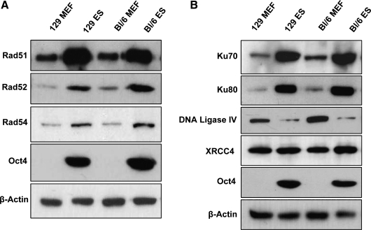
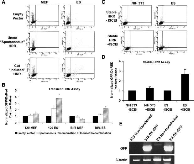
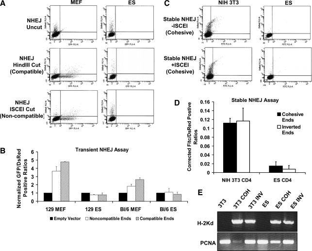
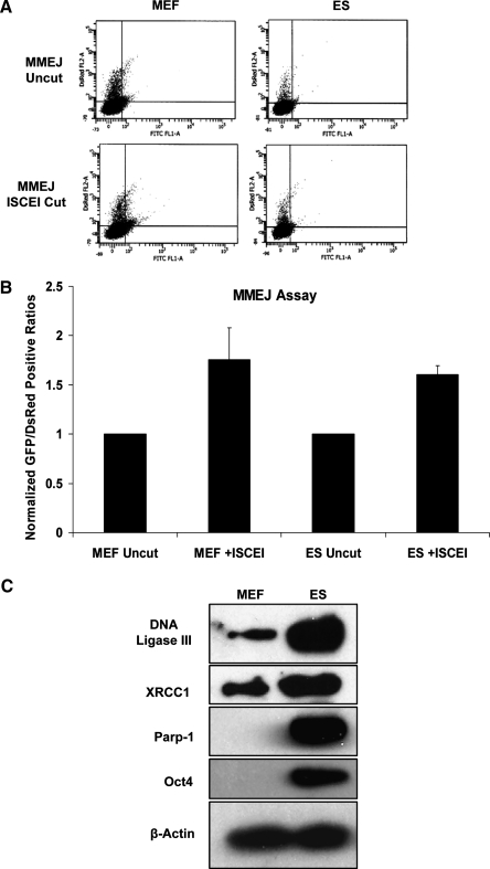
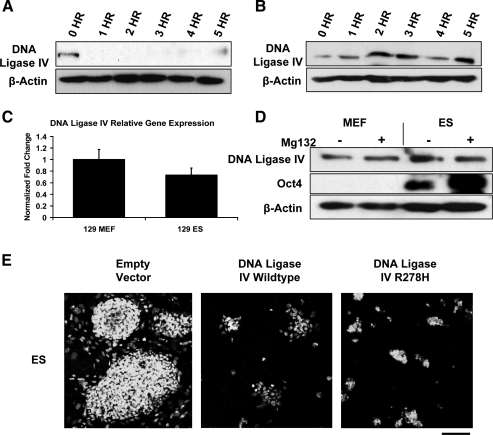
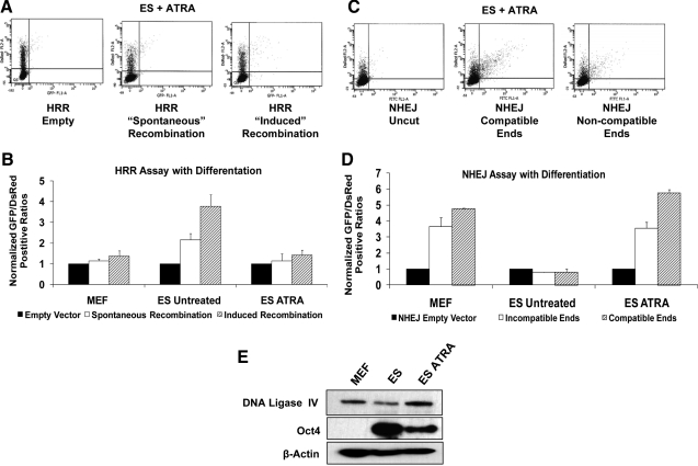
Embryonic stem (ES) cells give rise to all cell types of an organism. Since mutations at this embryonic stage would affect all cells and be detrimental to the overall health of an organism, robust mechanisms must exist to ensure that genomic integrity is maintained. To test this proposition, we compared the capacity of murine ES cells to repair DNA double-strand breaks with that of differentiated cells. Of the 2 major pathways that repair double-strand breaks, error-prone nonhomologous end joining (NHEJ) predominated in mouse embryonic fibroblasts, whereas the high fidelity homologous recombinational repair (HRR) predominated in ES cells. Microhomology-mediated end joining, an emerging repair pathway, persisted at low levels in all cell types examined. The levels of proteins involved in HRR and microhomology-mediated end joining were highly elevated in ES cells compared with mouse embryonic fibroblasts, whereas those for NHEJ were quite variable, with DNA Ligase IV expression low in ES cells. The half-life of DNA Ligase IV protein was also low in ES cells. Attempts to increase the abundance of DNA Ligase IV protein by overexpression or inhibition of its degradation, and thereby elevate NHEJ in ES cells, were unsuccessful. When ES cells were induced to differentiate, however, the level of DNA Ligase IV protein increased, as did the capacity to repair by NHEJ. The data suggest that preferential use of HRR rather than NHEJ may lend ES cells an additional layer of genomic protection and that the limited levels of DNA Ligase IV may account for the low level of NHEJ activity.


Figures

FIG. 1.
FIG. 1.
Basal protein expression of predominant DSB repair pathways. Western blots of whole cell lysates from either 129/SV or C57Bl/6 cells representing basal expression of several proteins involved in HRR (A) or NHEJ (B). Oct4 served as a marker of undifferentiated ES cells and β-actin was used as a loading control. DSB, double-strand break; HRR, homologous recombinational repair; NHEJ, nonhomologous end joining; ES cells, embryonic stem cells; MEF, mouse embryonic fibroblast.
FIG. 2.
FIG. 2.
Quantitation of HRR repair capacity. (A) Representative flow cytometry scatter plots for MEFs or ES cells derived from 129/Sv mice transiently transfected with pCAGGS (empty vector), circular pDR-GFP (spontaneous recombination), or I-SCE1-linearized pDR-GFP (induced recombination) and cotransfected with pDsRed to account for differences in transfection efficiency. Seventy-two hours posttransfection, cells were harvested and live sorted. A minimum of 10,000 cells were sorted per trial; only 5,000 are displayed. Axes represent mean fluorescent intensity on a 5-log scale. The X-axis represents GFP-positive cells; Y-axis is DsRed+ cells. pDR-GFP contains 2 nonfunctional copies of GFP separated by approximately 3 kb. Recombination between the 2 copies can yield functional GFP by gene conversion or a nonfunctional deletion product, which is not measured. Recombination between the 2 copies in the circular plasmid is noted as spontaneous recombination. When the I-SCE1 site located in one of the GFP copies is cut, a DSB in the plasmid occurs, which, if repaired, has induced recombination. (B) Quantitation of HRR activity in MEFs and ES cells from 129/Sv or C57Bl/6 stains. Data were generated based on the ratios of GFP+/DsRed+ cells and normalized to background caused from the empty vector from each cell type. A minimum of 10,000 cells per trial were used; 5,000 events are displayed. (C) Scatter plots of stably integrated DR-GFP in NIH 3T3 cells or 129/Sv ES cells. NIH 3T3 cells were used as a surrogate for primary MEFs, which cannot be selected for with their finite passage number. I-SCE1 expression vector pBaSCE or empty vector pCAGGS was introduced to induce site-specific DSBs and pDsRed was cotransfected for transfection efficiency. Seventy-two hours posttransfection, cells were harvested and sorted live. (D) Quantitation of HRR activity in stable cell lines represented in (C) before or after DSB induction. Data were collected as the total of GFP/DsRed+ cells and were normalized to respective cell lines transfected with empty vector. (E) PCR for GFP or β-actin on genomic DNA from nontransfected and stably transfected and pooled NIH 3T3 and ES cells to demonstrate pDR-GFP genomic integration. PCR, polymerase chain reaction.
FIG. 3.
FIG. 3.
NHEJ repair capacity in ES cells and differentiated cells. (A) Scatter plots for MEFs or ES cells from 129/Sv mouse strains transiently transfected with undigested (uncut/empty), HindIII-digested (compatible ends), or I-SCE1-digested (noncompatible ends) pEGFP-PEM1-AD2 vector and cotransfected with pDsRED. This vector contains a single GFP gene interrupted by a pem1 intron. Within the intron there is a killer ad2 exon, which prevents expression of functional GFP. Digestion with either HindIII or I-SCE1, located at the termini of the killer exon, allows for its removal. The pem1 intron can then be repaired by NHEJ and spliced out to render the GFP gene functional. Seventy-two hours posttransfection cells were harvested and analyzed by flow cytometry. At least 10,000 cells were examined per trial; 5,000 events are displayed. Axes are the same as described in Fig. 2. (B) Quantitation of NHEJ data from transiently transfected 129/Sv or C57Bl/6 MEFs and ES cells. Data were collected as total number of GFP+/DsRed+ cells and normalized to empty vector. (C) Representative scatter diagrams of NIH3T3 and 129/Sv ES cells stably transfected with pCOH-CD4 after transient transfection with pDsRed and pCAGGS or pBaSCE and staining with CD4-Fitc-conjugated antibody. (D) Cells were stably transfected with pCOH-CD4 (cohesive ends) or pINV-CD4 (inverted ends). Ends refer to the orientation of 2 I-SCE1 restriction sites within each constructs. Stable transfection leads to expression of H2Kd. When pBaSCE is introduced into stable cells, H2kd is cut out. Repair by NHEJ subsequently promotes expression of a downstream CD4 gene. Background CD4-positive cells from the pCAGGS empty vector were subtracted from total numbers after I-SCE1 introduction. (E) RNA was isolated from nontransfected or blasticidin-resistant pooled colonies of pCOH/pINV-transfected NIH 3T3 cells or 129/Sv ES cells. cDNA was synthesized and semiquantiative PCR was completed to demonstrate the stable integration of the constructs. PCR products were run on 0.8% agarose gels containing 3 μg/mL ethidium bromide. These cells normally do not express the cell surface marker H2Kd, but after transfection of pCOH or pINV, H2Kd expression was robust. PCNA demonstrated similar loading of pcr product per lane.
FIG. 4.
FIG. 4.
MMEJ repair capacity. (A) Whole cell lysates from 129/SV MEFs and ES cells were subjected to Western blotting using antibodies for DNA Ligase III, XRCC1, Parp1, Oct4, or β-actin. (B) Scatter diagrams for 129/Sv MEFs and ES cells transiently transfected with pDsRed and uncut or I-SCE1-linearized pCMV/I-SCE1/GFP vector and analyzed by flow cytometry 72 h posttransfection. Repair of this vector should only occur by MMEJ. This vector has a cassette inserted into the GFP gene, which renders it nonfunctional. Digestion with I-SCE1 and subsequent end processing to a repeat sequence at the ends of the cassette will render the GFP gene functional if ends are ligated together. (C) Quantitation of flow cytometric data. Data were accumulated from 10,000 cells per transfection as the total number of GFP+/DsRed+ cells and were normalized to the uncut vector for the respective cell line. MMEJ, microhomology-mediated end joining.
FIG. 5.
FIG. 5.
DNA Ligase IV regulation in mouse ES cells. 129/Sv ES cells (A) or MEFs (B) were treated with 25 μg/mL of cycloheximide to block new protein synthesis or left untreated and harvested at the specified time points for Western blotting with DNA Ligase IV antibody. β-actin served as a loading control. (C) RNA from untreated 129/Sv MEFs and ES cells was isolated and subjected to semiquantitative PCR. Data are graphed as a ratio of DNA Ligase IV/Gapdh. (D) 129/Sv MEFs and ES cells were treated with 30 μM of the proteasome inhibitor MG-132 or left untreated and harvested after 4 h. Western blotting was performed using antibodies against DNA Ligase IV or Oct4 to demonstrate undifferentiated ES cells. β-actin served as a loading control. (E) 129/Sv ES cells were transfected with GFP, GFP-DNA Ligase IV wild type, and GFP-DNA Ligase IV hypomorphic mutant (R278H), and allowed to grow for 72 h. Nuclei were stained with Draq5. While large ES cell colonies are visible in the vector-only-transfected cells, the GFP DNA Ligase IV wild-type and mutant-transfected cells were much fewer in number. Scale bar = 100 μm.
FIG. 6.
FIG. 6.
Differentiated ES cell DSB repair patterns. ES cells were differentiated by treatment with ATRA. (A) Scatter diagrams of transiently transfected differentiated ES cells using vectors pCAGGS and pDR-GFP (A) or pEGFP-PEM1-AD2 (C). (B) Flow cytometry quantitation of differentiated ES cell capacity to repair DSBs by HRR (B) or NHEJ (D) as compared with MEFs or undifferentiated ES cells. Data accumulation and methodology is identical to that of Figs. 3B and 4B. (E) Whole-cell lysates from MEFs, undifferentiated ES cells, or ATRA-treated ES cells were subjected to Western blotting using antibodies to DNA Ligase IV, Oct4, and β-actin. DNA Ligase IV is increased in differentiated cells. Oct4 is still present in these cells, suggesting that complete differentiation has not yet occurred. ATRA, all-trans retinoic acid.

References

    1. Friedberg EC. Meira LB. Database of mouse strains carrying targeted mutations in genes affecting biological responses to DNA damage Version 7. DNA Repair (Amst) 2006;5:189–209. - PubMed
    1. Cervantes RB. Stringer JR. Shao C. Tischfield JA. Stambrook PJ. Embryonic stem cells and somatic cells differ in mutation frequency and type. Proc Natl Acad Sci USA. 2002;99:3586–3590. - PMC - PubMed
    1. Savatier P. Huang S. Szekely L. Wiman KG. Samarut J. Contrasting patterns of retinoblastoma protein expression in mouse embryonic stem cells and embryonic fibroblasts. Oncogene. 1994;9:809–818. - PubMed
    1. Savatier P. Lapillonne H. Jirmanova L. Vitelli L. Samarut J. Analysis of the cell cycle in mouse embryonic stem cells. Methods Mol Biol. 2002;185:27–33. - PubMed
    1. Hong Y. Stambrook PJ. Restoration of an absent G1 arrest and protection from apoptosis in embryonic stem cells after ionizing radiation. Proc Natl Acad Sci USA. 2004;101:14443–14448. - PMC - PubMed

Publication types