. 2010 Jun 22;119(7):273-82.
doi: 10.1042/CS20100084.

Angiotensin II and the ERK pathway mediate the induction of myocardin by hypoxia in cultured rat neonatal cardiomyocytes

Affiliations

Angiotensin II and the ERK pathway mediate the induction of myocardin by hypoxia in cultured rat neonatal cardiomyocytes

Chiung-Zuan Chiu et al. Clin Sci (Lond). .

Abstract

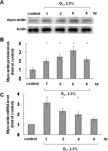
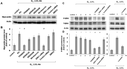
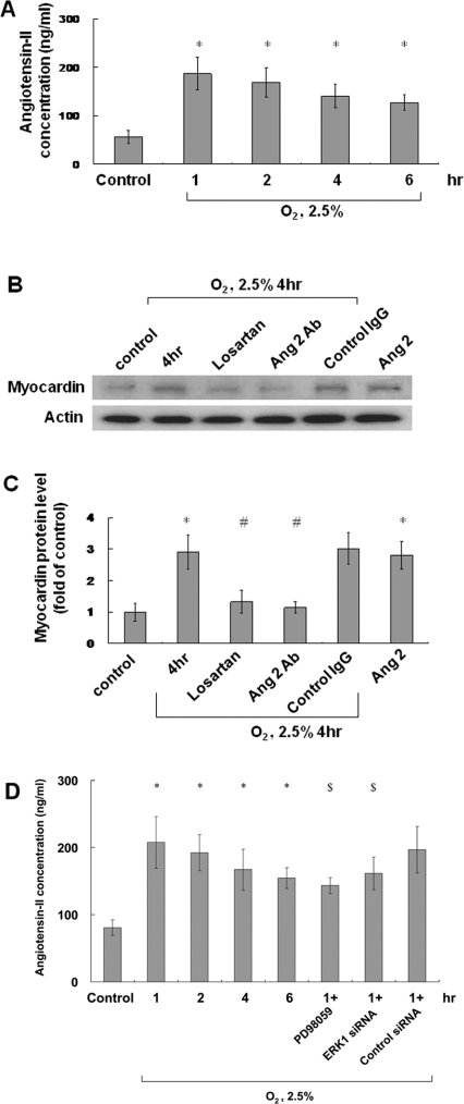
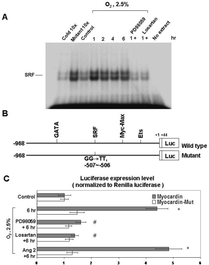
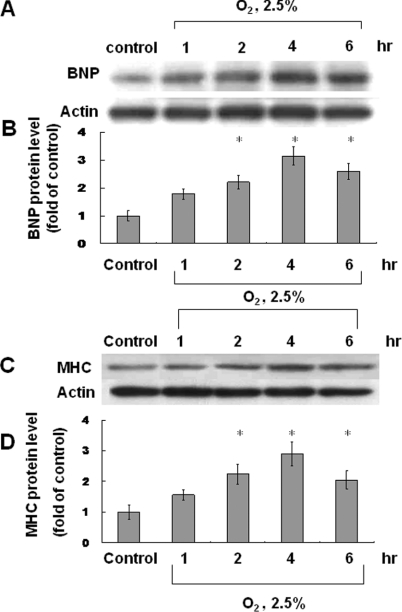
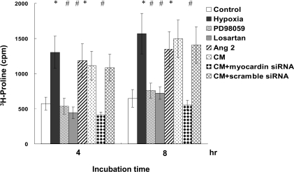
Hypoxic injury to cardiomyocytes is a stress that causes cardiac pathology through cardiac-restricted gene expression. SRF (serum-response factor) and myocardin are important for cardiomyocyte growth and differentiation in response to myocardial injuries. Previous studies have indicated that AngII (angiotensin II) stimulates both myocardin expression and cardiomyocyte hypertrophy. In the present study, we evaluated the expression of myocardin and AngII after hypoxia in regulating gene transcription in neonatal cardiomyocytes. Cultured rat neonatal cardiomyocytes were subjected to hypoxia, and the expression of myocardin and AngII were evaluated. Different signal transduction pathway inhibitors were used to identify the pathway(s) responsible for myocardin expression. An EMSA (electrophoretic mobility-shift assay) was used to identify myocardin/SRF binding, and a luciferase assay was used to identify transcriptional activity of myocardin/SRF in neonatal cardiomyocytes. Both myocardin and AngII expression increased after hypoxia, with AngII appearing at an earlier time point than myocardin. Myocardin expression was stimulated by AngII and ERK (extracellular-signal-regulated kinase) phosphorylation, but was suppressed by an ARB (AngII type 1 receptor blocker), an ERK pathway inhibitor and myocardin siRNA (small interfering RNA). AngII increased both myocardin expression and transcription in neonatal cardiomyocytes. Binding of myocardin/SRF was identified using an EMSA, and a luciferase assay indicated the transcription of myocardin/SRF in neonatal cardiomyocytes. Increased BNP (B-type natriuretic peptide), MHC (myosin heavy chain) and [(3)H]proline incorporation into cardiomyocytes was identified after hypoxia with the presence of myocardin in hypertrophic cardiomyocytes. In conclusion, hypoxia in cardiomyocytes increased myocardin expression, which is mediated by the induction of AngII and the ERK pathway, to cause cardiomyocyte hypertrophy. Myocardial hypertrophy was identified as an increase in transcriptional activities, elevated hypertrophic and cardiomyocyte phenotype markers, and morphological hypertrophic changes in cardiomyocytes.


Figures

Figure 1
Figure 1. Effect of hypoxia on myocardin protein and mRNA levels
Neonatal cardiomyocytes were subjected to hypoxia for 1–6 h, and total cell lysates were immunoblotted with an anti-myocardin antibody (A) or (C) extracted to determine mRNA levels. (A and B) Myocardin protein level increased and reached a peak after 4 h of hypoxia. Actin is used to show equal amounts of protein loading in each lane. (C) Myocardin mRNA levels reached a peak after 1 h of hypoxia and then declined. *P<0.01 compared with normoxia control (n=3).
Figure 2
Figure 2. Effect of signalling pathway inhibitors on hypoxia-induced myocardin expression and ERK phosphorylation
(A and B) ERK pathway mediates hypoxia-induced myocardin expression in neonatal cardiomyocytes. Neonatal cardiomyocytes were pre-treated with an ERK pathway inhibitor (PD98059), a JNK inhibitor (SP600125), a p38 MAPK inhibitor (SB203580), a PI3K/Akt inhibitor (wortmannin) or ERK siRNA, followed by hypoxia for 4 h. Neonatal cardiomyocytes were harvested and cell lysates were analysed by Western blotting using an anti-myocardin antibody. Result are normalized to actin levels. *P<0.01 compared with normoxia control; #P<0.01 compared with hypoxia for 4 h (n=3). (C and D) Hypoxia-induced phosphorylation of ERK in neonatal cardiomyocytes. Neonatal cardiomyocytes were subjected to normoxia or hypoxia for 1–6 h in the presence or absence of inhibitors, and cell lysates were collected for Western blot analysis using antibodies against total ERK (T-ERK) and phospho-ERK (P-ERK). *P<0.01 compared with normoxia control; #P<0.01 compared with hypoxia for 1 h (n=3).
Figure 3
Figure 3. Hypoxia induces AngII expression and mediates hypoxia-induced myocardin expression
(A) AngII was measured in cell lysates and the culture medium by a quantitative competitive ELISA, using a specific anti-AngII antibody, following 1–6 h of hypoxia. (B and C) Neonatal cardiomyocytes were subjected to normoxia or hypoxia for 4 h in the absence or presence of losartan (100 nmol/l) or an anti-AngII antibody and the addition of exogenous AngII (10 nmol/l). Total cell lysates were immunoblotted with an anti-myocardin antibody. Actin is used to show equal amounts of protein loading in each lane. (D) Hypoxia-induced AngII expression was suppressed by PD98059 and ERK siRNA. *P<0.01 compared with normoxia control; #P<0.01 compared with 4 h of hypoxia; $P<0.05 compared with 1 h of hypoxia (n=3).
Figure 4
Figure 4. Hypoxia induces myocardin/SFR binding (A) and myocardin transcriptional activity (B and C) in cardiomyocytes
(A) Increased binding between myocardin and SRF under 2.5% O2 as determined using an EMSA. The binding between myocardin and SRF was suppressed by PD98059 and losartan. (B) Wild-type and myocardin mutant used for the luciferase assay. (C) Hypoxia (2.5% O2) increased the transcriptional activity of myocardin when compared with the myocardin mutant (Mut) as determined using a luciferase reporter assay. The transcriptional activity was suppressed by PD98059 and losartan. Addition of exogenous AngII (10 nmol/l) increased the transcriptional activity in neonatal cardiomyocyte similar to that of 2.5% O2. *P<0.01 compared with normoxia control; #P<0.01 compared with 6 h (n=3).
Figure 5
Figure 5. Hypoxia induces the expression of BNP and MHC in cardiomyocytes
BNP (A and B) and MHC (C and D) protein levels were both increased after hypoxia (2.5% O2), reaching peak levels after 4 h.*P<0.01 compared with normoxia control (n=3).
Figure 6
Figure 6. Hypoxia induces protein synthesis in neonatal cardiomyocytes
Incorporation of [3H]proline into neonatal cardiomyocytes increased after hypoxia and the exogenous addition of AngII for 4–8 h, and was suppressed by PD98059, losartan, and myocardin siRNA. *P<0.01 compared with normoxia control; #P<0.01 compared with hypoxia (n=6). CM, conditioned medium.

References

    1. Miano J. M. Serum response factor: toggling between disparate programs of gene expression. J. Mol. Cell. Cardiol. 2003;35:577–593. - PubMed
    1. Wang D., Chang P. S., Wang Z., Sutherland L., Richardson J. A., Small E., Krieg P. A., Olson E. N. Activation of cardiac gene expression by myocardin, a transcriptional cofactor for serum response factor. Cell. 2001;105:851–862. - PubMed
    1. Zhang X., Azhar G., Zhong Y., Wei J. Y. Identification of a novel serum response factor cofactor in cardiac gene regulation. J. Biol. Chem. 2004;279:55626–55632. - PubMed
    1. Wang D. Z., Li S., Hockemeyer D., Sutherland L., Wang Z., Schratt G. Potentiation of serum response factor activity by a family of myocardin-related transcription factors. Proc. Natl. Acad. Sci. U.S.A. 2002;99:14855–14860. - PMC - PubMed
    1. Kontaraji J. E., Parthenakis F. I., Patrianakos A. P., Karalis I. K., Vardas P. E. Altered expression of early marker genes in circulating cells of patients with hypertrophic cardiomyopathy. Cardiovasc. Pathol. 2007;16:329–335. - PubMed

MeSH terms