Facial reconstruction--anatomical art or artistic anatomy?
- PMID: 20447245
- PMCID: PMC2815945
- DOI: 10.1111/j.1469-7580.2009.01182.x
Facial reconstruction--anatomical art or artistic anatomy?
Abstract
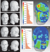
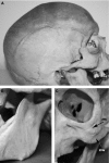
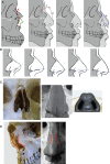
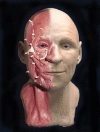
Facial reconstruction is employed in the context of forensic investigation and for creating three-dimensional portraits of people from the past, from ancient Egyptian mummies and bog bodies to digital animations of J. S. Bach. This paper considers a facial reconstruction method (commonly known as the Manchester method) associated with the depiction and identification of the deceased from skeletal remains. Issues of artistic licence and scientific rigour, in relation to soft tissue reconstruction, anatomical variation and skeletal assessment, are discussed. The need for artistic interpretation is greatest where only skeletal material is available, particularly for the morphology of the ears and mouth, and with the skin for an ageing adult. The greatest accuracy is possible when information is available from preserved soft tissue, from a portrait, or from a pathological condition or healed injury.
Figures


Similar articles
-
Superimposition and reconstruction in forensic facial identification: a survey.Forensic Sci Int. 1995 Oct 30;75(2-3):101-20. doi: 10.1016/0379-0738(95)01770-4. Forensic Sci Int. 1995. PMID: 8586334 Review.
-
Facial approximation of 'angel': case specific methodological review.Forensic Sci Int. 2014 Apr;237:e30-41. doi: 10.1016/j.forsciint.2013.12.039. Epub 2014 Jan 28. Forensic Sci Int. 2014. PMID: 24582078
-
Soft tissue thickness in Brazilian adults of different skeletal classes and facial types: A cone beam CT - Study.Leg Med (Tokyo). 2020 Nov;47:101743. doi: 10.1016/j.legalmed.2020.101743. Epub 2020 Jul 3. Leg Med (Tokyo). 2020. PMID: 32659706
-
Pilot study of facial soft tissue thickness differences among three skeletal classes in Japanese females.Forensic Sci Int. 2010 Feb 25;195(1-3):165.e1-5. doi: 10.1016/j.forsciint.2009.10.013. Epub 2009 Nov 25. Forensic Sci Int. 2010. PMID: 19942386
-
Forensic three-dimensional facial reconstruction: historical review and contemporary developments.J Forensic Sci. 1997 Jul;42(4):653-61. J Forensic Sci. 1997. PMID: 9243827 Review.
Cited by
-
Comparative Analysis of Korean Nasal Morphology Using Cone-Beam Computed Tomography.Healthcare (Basel). 2024 Sep 13;12(18):1839. doi: 10.3390/healthcare12181839. Healthcare (Basel). 2024. PMID: 39337180 Free PMC article.
-
A method for automatic forensic facial reconstruction based on dense statistics of soft tissue thickness.PLoS One. 2019 Jan 23;14(1):e0210257. doi: 10.1371/journal.pone.0210257. eCollection 2019. PLoS One. 2019. PMID: 30673719 Free PMC article.
-
Craniofacial anthropometric investigation of relationships between the nose and nasal aperture using 3D computed tomography of Korean subjects.Sci Rep. 2020 Sep 30;10(1):16077. doi: 10.1038/s41598-020-73127-8. Sci Rep. 2020. PMID: 32999371 Free PMC article.
-
Comparison of Nasal Dimensions According to the Facial and Nasal Indices Using Cone-Beam Computed Tomography.J Pers Med. 2024 Apr 14;14(4):415. doi: 10.3390/jpm14040415. J Pers Med. 2024. PMID: 38673042 Free PMC article.
-
"Bochdalek's" skull: morphology report and reconstruction of face.Forensic Sci Med Pathol. 2012 Dec;8(4):451-9. doi: 10.1007/s12024-012-9375-5. Epub 2012 Aug 24. Forensic Sci Med Pathol. 2012. PMID: 22918853
References
-
- Algemeen Vader Maasmeisje overleden in gevangenis. 2009. Published 12 May 2009. Available at: http://translate.google.co.uk/translate?hl=enandsl=nlandu=http://www.nu.....
-
- Angel JL. Proceedings of Meetings of American Academy of Forensic Science. St. Louis, MO: 1978. Restoration of head and face for identification.
-
- Asingh P, Lynnerup N. Grauballe Man – An Iron Age Bog Body Revisited. Jutland Archaeological Society: Moesgaard; 2007.
-
- Aufderheide AC. The Scientific Study of Mummies. Cambridge University Press: Cambridge; 2003.
-
- Auslebrook WA, Becker PJ, Iscan MY. Facial soft tissue thicknesses in the adult male Zulu. Forensic Sci Int. 1996;79:83–102. - PubMed
Publication types
MeSH terms
LinkOut - more resources
Full Text Sources
Miscellaneous

