. 2010 Apr;216(4):446-62.
doi: 10.1111/j.1469-7580.2009.01209.x.

Muscle moment arms of the gibbon hind limb: implications for hylobatid locomotion

Affiliations

Muscle moment arms of the gibbon hind limb: implications for hylobatid locomotion

Anthony J Channon et al. J Anat. 2010 Apr.

Abstract

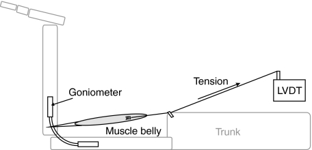
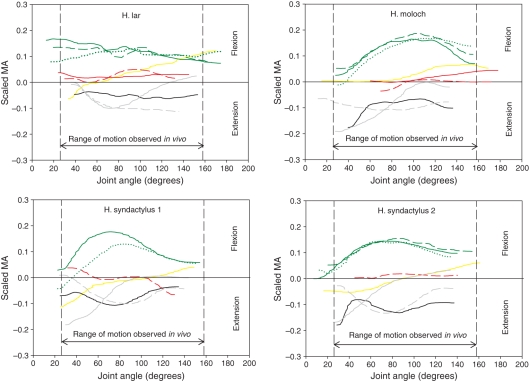
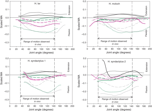
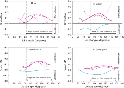
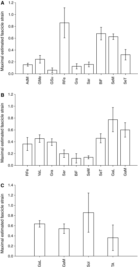
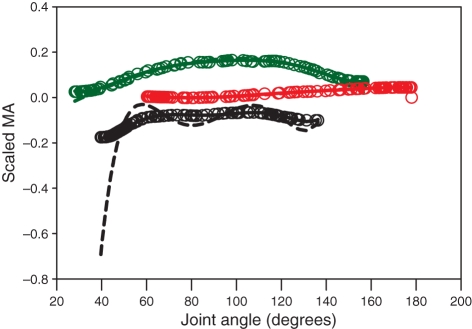
Muscles facilitate skeletal movement via the production of a torque or moment about a joint. The magnitude of the moment produced depends on both the force of muscular contraction and the size of the moment arm used to rotate the joint. Hence, larger muscle moment arms generate larger joint torques and forces at the point of application. The moment arms of a number of gibbon hind limb muscles were measured on four cadaveric specimens (one Hylobates lar, one H. moloch and two H. syndactylus). The tendon travel technique was used, utilizing an electro-goniometer and a linear voltage displacement transducer. The data were analysed using a technique based on a differentiated cubic spline and normalized to remove the effect of body size. The data demonstrated a functional differentiation between voluminous muscles with short fascicles having small muscle moment arms and muscles with longer fascicles and comparatively smaller physiological cross-sectional area having longer muscle moment arms. The functional implications of these particular configurations were simulated using a simple geometric fascicle strain model that predicts that the rectus femoris and gastrocnemius muscles are more likely to act primarily at their distal joints (knee and ankle, respectively) because they have short fascicles. The data also show that the main hip and knee extensors maintain a very small moment arm throughout the range of joint angles seen in the locomotion of gibbons, which (coupled to voluminous, short-fascicled muscles) might help facilitate rapid joint rotation during powerful movements.


Figures

Fig. 2
Fig. 2
The joint angles used in the analyses.
Fig. 1
Fig. 1
The apparatus used for data collection. LVDT, Linear voltage displacement transducer.
Fig. 3
Fig. 3
Decreasing residual sum of squares (RSS) values for increasing orders of polynomial used to fit data from the rectus femoris at the hip. The filled bar represents the least complex model, which has an RSS of less than the 5% threshold.
Fig. 4
Fig. 4
Scaled moment arms (MAs) at the hip for the four specimens. Solid yellow line, adductor magnus; solid red line, gluteus medius; dashed red line, gluteus superficialis; solid black line, rectus femoris; solid grey line, gracilis; dashed grey line, sartorius; solid green line, biceps femoris; dashed green line, semimembranosus; dotted green line, semitendinosus. The vertical black dashed lines indicate the range of joint motion used during jumping (see Materials and methods for jumping data collection).
Fig. 5
Fig. 5
Scaled moment arms (MAs) at the knee for the four specimens. Solid black line, rectus femoris; dotted black line, vastus lateralis; solid grey line, gracilis; dashed grey line, sartorius; solid green line, biceps femoris; dashed green line, semimembranosus; dotted green line, semitendinosus; solid pink line, gastrocnemius lateralis; dashed pink line, gastrocnemius medialis. The vertical black dashed lines indicate the range of joint motion used during jumping (see Materials and methods for jumping data collection).
Fig. 6
Fig. 6
Scaled moment arms (MAs) at the ankle for the four specimens. Solid pink line, gastrocnemius lateralis; dashed pink line, gastrocnemius medialis; dotted pink line, soleus; cyan line, tibialis anterior. The vertical black dashed lines indicate the range of joint motion used during jumping (see Materials and methods for jumping data collection).
Fig. 7
Fig. 7
Estimated fascicle strain during the range of motion tested for muscles at the hip (A), knee (B) and ankle (C). AdM, adductor magnus; BiF, biceps femoris; SeM, semimembranosus; SeT, semitendinosus; Sol, soleus; GMe, gluteus medius; GSu, gluteus superficialis; Gra, gracilis; Sar, sartorius; RFe, rectus femoris; VaL, vastus lateralis; GaL, gastrocnemius lateralis; GaM, gastrocnemius medialis; TiA, tibialis anterior.
Fig. 8
Fig. 8
The differences in predicted moment arm (MA) for polynomial-based and spline-based techniques for three muscles. Black, rectus femoris muscle; green, biceps femoris muscle; red, gluteus medius muscle. Dashed lines, traditional (polynomial-based) technique; open circles, spline based technique; solid lines, polynomial fitted to the splined data.

Similar articles

Cited by

References

    1. Ackland DC, Pandy MG. Lines of action and stabilizing potential of the shoulder musculature. J Anat. 2009;215:184–197. - PMC - PubMed
    1. Alexander RM. Allometry of the limbs of antelopes (Bovidae) J Zool. 1977;183:125–146.
    1. Alexander RM. Elastic energy storage in running vertebrates. Amer Zool. 1984;24:85–94.
    1. Alexander RM. Leg design and jumping technique for humans, other vertebrates and insects. Philos Trans R Soc Lond B Biol Sci. 1995;347:235–248. - PubMed
    1. Alexander RM. Principles of Animal Locomotion. Princeton: Princeton University Press; 1996.

Publication types

LinkOut - more resources