. 2010 May;11(3):335-46.
doi: 10.1111/j.1364-3703.2009.00606.x.

Helper component-proteinase (HC-Pro) protein of Papaya ringspot virus interacts with papaya calreticulin

Affiliations

Helper component-proteinase (HC-Pro) protein of Papaya ringspot virus interacts with papaya calreticulin

Wentao Shen et al. Mol Plant Pathol. 2010 May.

Abstract

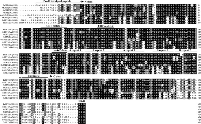
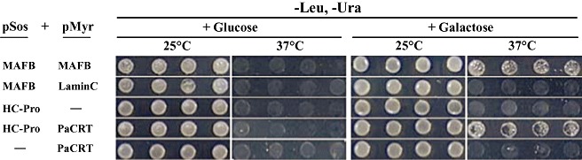
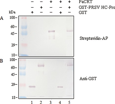
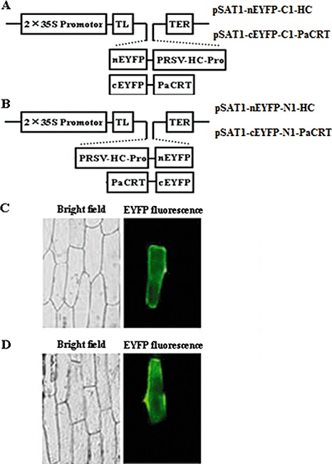
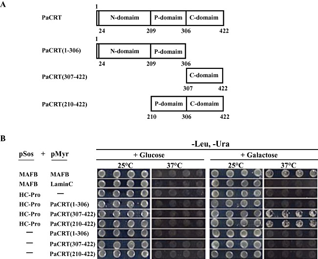
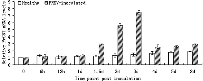
Potyviral helper component-proteinase (HC-Pro) is a multifunctional protein involved in plant-virus interactions. In this study, we constructed a Carica papaya L. plant cDNA library to investigate the host factors interacting with Papaya ringspot virus (PRSV) HC-Pro using a Sos recruitment two-hybrid system (SRS). We confirmed that the full-length papaya calreticulin, designated PaCRT (GenBank accession no. FJ913889), interacts specifically with PRSV HC-Pro in yeast, in vitro and in plant cells using SRS, in vitro protein-binding assay and bimolecular fluorescent complementation assay, respectively. SRS analysis of the interaction between three PaCRT deletion mutants and PRSV HC-Pro demonstrated that the C-domain (residues 307-422), with a high Ca(2+)-binding capacity, was responsible for binding to PRSV HC-Pro. In addition, quantitative real-time reverse transcriptase-polymerase chain reaction assay showed that the expression of PaCRT mRNA was significantly upregulated in the primary stage of PRSV infection, and decreased to near-basal expression levels in noninoculated (healthy) papaya plants with virus accumulation inside host cells. PaCRT is a new calcium-binding protein that interacts with potyviral HC-Pro. It is proposed that the upregulated expression of PaCRT mRNA may be an early defence-related response to PRSV infection in the host plant, and that interaction between PRSV HC-Pro and PaCRT may be involved in plant calcium signalling pathways which could interfere with virus infection or host defence.


Figures

Figure 1
Figure 1
Amino acid sequence alignment and domains of Carica papaya calreticulin (PaCRT). Sequence alignment of CRT proteins from Carica papaya (Pa; database accession number ACQ91203), Arabidopsis thaliana (At; CRT1: AAC49695; CRT2: NP172392; CRT3: AAC49697), Oryza sativa (Os; OsCRT1/2: BAA88900; CRT3: BAC06263), Nicotiana tabacum (Nt; CAA59694) and Triticum aestivum (Ta; EF452301) was performed using the sequence manipulation suite (Paul Stothard, University of Alberta, Edmonton, AB, Canada). Residues that were identical in all of these proteins are highlighted on a black background. The arrows indicate the approximate positions of the three domains (N, P and C). The predicted signal peptide, conserved CRT family motifs (1, KHEQKLDCGGGYVKLL; 2, IMFGPDICG) and triplicate repeats (A, PXXIXDPXX KKPEXWDD; B, GXWXAXXIXNPXYK) are underlined (Michalak et al. 1999). The putative nuclear targeting sequence (PPKKIKDPE) is marked with a bold underline. The endoplasmic reticulum retention sequence HDEL is indicated at the C‐terminus.
Figure 2
Figure 2
Detection of the interaction between Papaya ringspot virus helper component‐proteinase (PRSV HC‐Pro) and Carica papaya calreticulin (PaCRT) by the Sos recruitment assay. Saccharomyces cerevisiae strain cdc25H was transformed with the indicated plasmid combinations. Four colonies from each transformant were picked up, resuspended and diluted to optical densities at 600 nm of 0.5 in sterile water. An aliquot of 2.5 µL of each dilution was patched in rows onto each of two synthetic glucose minimal medium without leucine and uracil [SD/glucose (−UL)] and two synthetic galactose minimal medium without leucine and uracil [SD/galactose (−UL)] plates, and one of each type of plate was incubated at the permissive temperature or nonpermissive temperature (25 or 37°C) for 5 days to compare the growth of yeast. pMyr‐MAFB and pSos‐MAFB were used as positive controls, and pMyr‐Lamin C and pSos‐MAFB were used as negative controls.
Figure 3
Figure 3
In vitro protein‐binding assay of Papaya ringspot virus helper component‐proteinase (PRSV HC‐Pro) and Carica papaya calreticulin (PaCRT). In vitro translated biotinylated PaCRT was incubated with purified glutathione S‐transferase (GST) or GST‐PRSV HC‐Pro, which was immobilized on MagneGST particles. After incubation, the proteins interacting with GST or GST‐PRSV HC‐Pro were bound to the MagneGST particles. The eluted GST‐bound proteins (lane 4) or GST‐PRSV HC‐Pro‐bound proteins (lane 5) were then analysed by 10% sodium dodecylsulphate‐polyacrylamide gel electrophoresis (SDS‐PAGE) and probed with streptavidin‐alkaline phosphatase (streptavidin‐AP) (A) or anti‐GST antibody (B) (GST, 26.0 kDa; GST‐PRSV HC‐Pro, 78.0 kDa; biotinylated PaCRT, 48.2 kDa). The purified GST (lane 1), purified GST‐PRSV HC‐Pro (lane 2) and in vitro translated biotinylated PaCRT (lane 3) were used as controls, respectively. The prestained dual colour protein size marker (Tiangen) is indicated on the left of the panel.
Figure 4
Figure 4
Interaction between Papaya ringspot virus helper component‐proteinase (PRSV HC‐Pro) and Carica papaya calreticulin (PaCRT) by bimolecular fluorescent complementation (BiFC) assay in living plant cells. (A) Schematic structure of pSAT1‐nEYFP‐C1‐HC and pSAT1‐cEYFP‐C1‐PaCRT constructs used for BiFC assay. (B) Schematic structure of pSAT1‐nEYFP‐N1‐HC and pSAT1‐cEYFP‐N1‐PaCRT constructs used for BiFC assay; 2 × 35S, tandem cauliflower mosaic virus (CaMV) 35S promoter; TL, Tobacco etch virus (TEV) translation leader; TER, CaMV 35S poly(A) transcriptional terminator; nEYFP and cEYFP, N‐terminal and C‐terminal fragments of yellow fluorescent protein variant derived from enhanced green fluorescent protein (EYFP); C1 and N1 vectors produce fusion to the C‐ and N‐termini of cEYFP and nEYFP, respectively. (C) The reconstructed YFP signal detected in onion (Allium cepa) epidermal cells by epifluorescence microscopy after bombardment with (A) constructs. (D) The reconstructed YFP signal detected in onion epidermal cells by epifluorescence microscopy after bombardment with (B) constructs. Left panels, bright field; right panels, EYFP fluorescence (green).
Figure 5
Figure 5
Schematic representation of Carica papaya calreticulin (PaCRT) mutants and identification of domains of PaCRT necessary for interaction with Papaya ringspot virus helper component‐proteinase (PRSV HC‐Pro). (A) Schematic representation of PaCRT and three PaCRT mutants: PaCRT, PaCRT(1–306), PaCRT(307–422) and PaCRT(210–422). (B) Interaction of PRSV HC‐Pro and mutants of PaCRT in Saccharomyces cerevisiae strain cdc25H cells. Four colonies transformed with the indicated plasmid combinations (B) from each transformant were picked up, resuspended and diluted to optical densities at 600 nm of 0.5 in sterile water. An aliquot of 2.5 µL of each dilution was patched in rows onto each of two synthetic glucose minimal medium without leucine and uracil [SD/glucose (−UL)] and two synthetic galactose minimal medium without leucine and uracil [SD/galactose (−UL)] plates, and one of each type of plate was incubated at the permissive temperature or nonpermissive temperature (25 or 37°C) for 5 days to compare the growth of yeast. pMyr‐MAFB and pSos‐MAFB were used as positive controls, and pMyr‐Lamin C and pSos‐MAFB were used as negative controls.
Figure 6
Figure 6
Comparison of relative Carica papaya calreticulin (PaCRT) mRNA levels in healthy and PRSV‐inoculated Carica papaya plants by quantitative real‐time reverse transcriptasepolymerase chain reaction (qRT‐PCR). Data shown are expressed as the mean ± SD of three independent samples (P < 0.05).
Figure 7
Figure 7
Phylogenetic analysis of calreticulin (CRT) protein sequences in plants and two mammalians. Sequence data for CRT protein were obtained from the GenBank database and the accession numbers are indicated in parentheses. The phylogenetic analysis was performed using Geneious Pro 4.5 software (Biomatters, Auckland, New Zealand). Two distinct groups and two plant subgroups are indicated to the left of the phylogenetic tree.

Similar articles

Cited by

References

    1. Ahlquist, P. , Noueiry, A.O. , Lee, W.M. , Kushner, D.B. and Dye, B.T. (2003) Host factors in positive‐strand RNA virus genome replication. J. Virol. 77, 8181–8186. - PMC - PubMed
    1. Anandalakshmi, R. , Marathe, R. , Ge, X. , Herr, J.M., Jr , Mau, C. , Mallory, A. , Pruss, G. , Bowman, L. and Vance, V.B. (2000) A calmodulin‐related protein that suppresses posttranscriptional gene silencing in plants. Science, 290, 142–144. - PubMed
    1. Ballut, L. , Drucker, M. , Pugnière, M. , Cambon, F. , Blanc, S. , Roquet, F. , Candresse, T. , Schmid, H.P. , Nicolas, P. , Gall, O.L. and Badaoui, S. (2005) HcPro, a multifunctional protein encoded by a plant RNA virus, targets the 20S proteasome and affects its enzymic activities. J. Gen. Virol. 86, 2595–2603. - PubMed
    1. Bau, H.J. , Cheng, Y.H. , Yu, T.A. , Yang, J.S. and Yeh, S.D. (2003) Broad‐spectrum resistance to different geographic strains of papaya ringspot virus in coat protein gene transgenic papaya. Phytopathology 93, 112–120. - PubMed
    1. Bracha‐Drori, K. , Shichrur, K. , Katz, A. , Oliva, M. , Angelovici, R. , Yalovsky, S. and Ohad, N. (2004) Detection of protein–protein interactions in plants using bimolecular fluorescence complementation. Plant J. 40, 419–427. - PubMed

Publication types

MeSH terms