. 2010 Jun;163(6):630-7.
doi: 10.1016/j.chemphyslip.2010.04.011. Epub 2010 May 4.

Structure and water permeability of fully hydrated diphytanoylPC

Affiliations

Structure and water permeability of fully hydrated diphytanoylPC

Stephanie Tristram-Nagle et al. Chem Phys Lipids. 2010 Jun.

Abstract

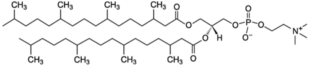
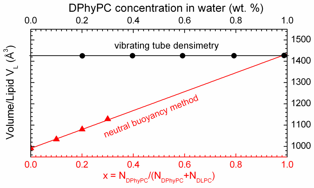
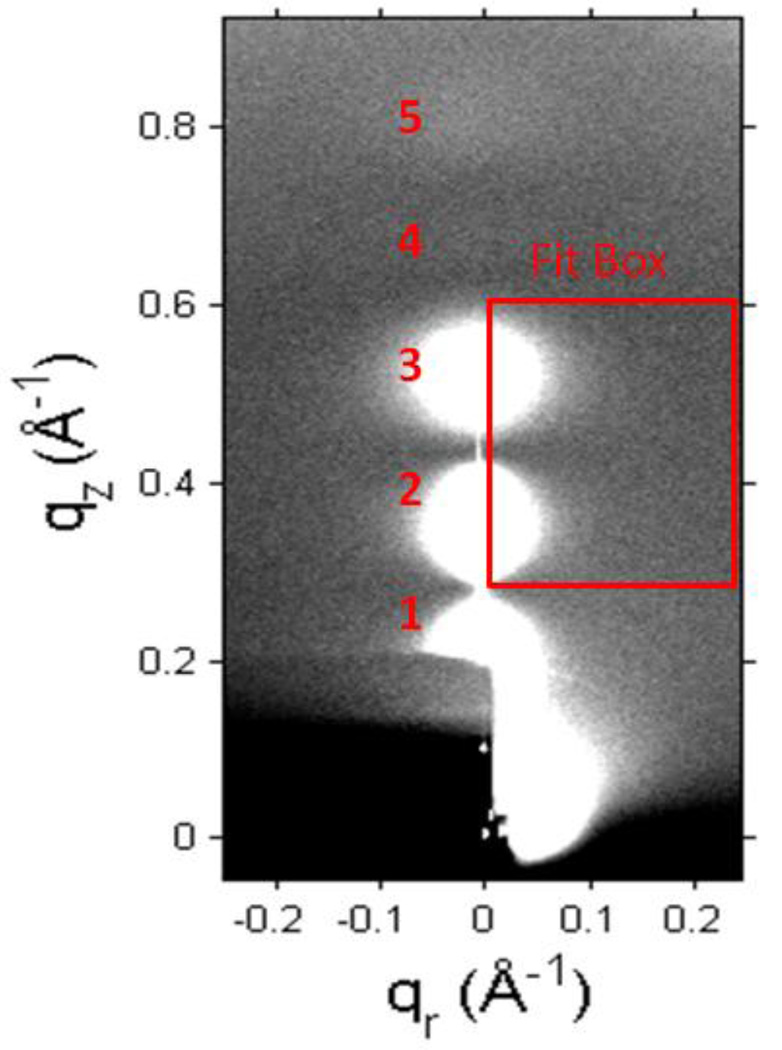
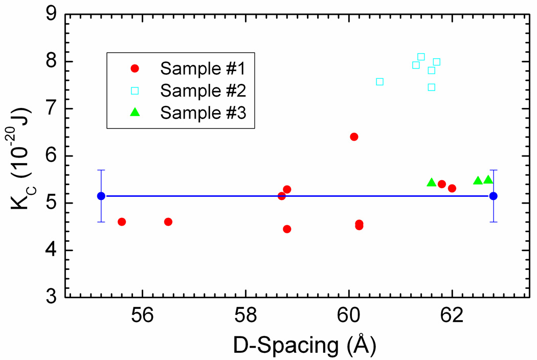
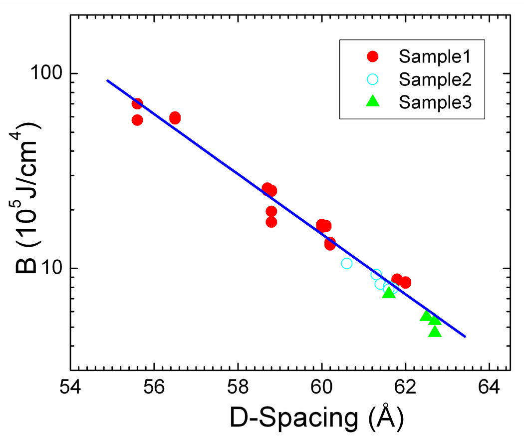
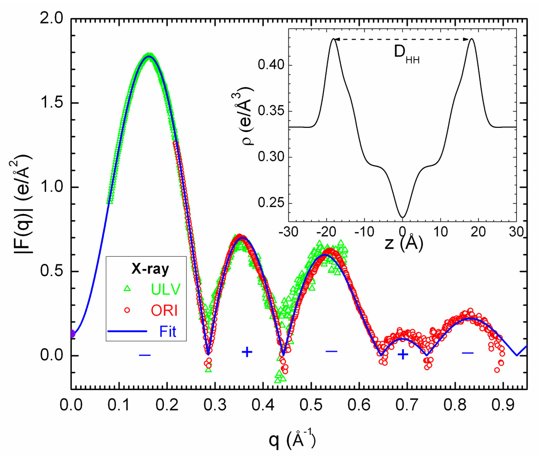
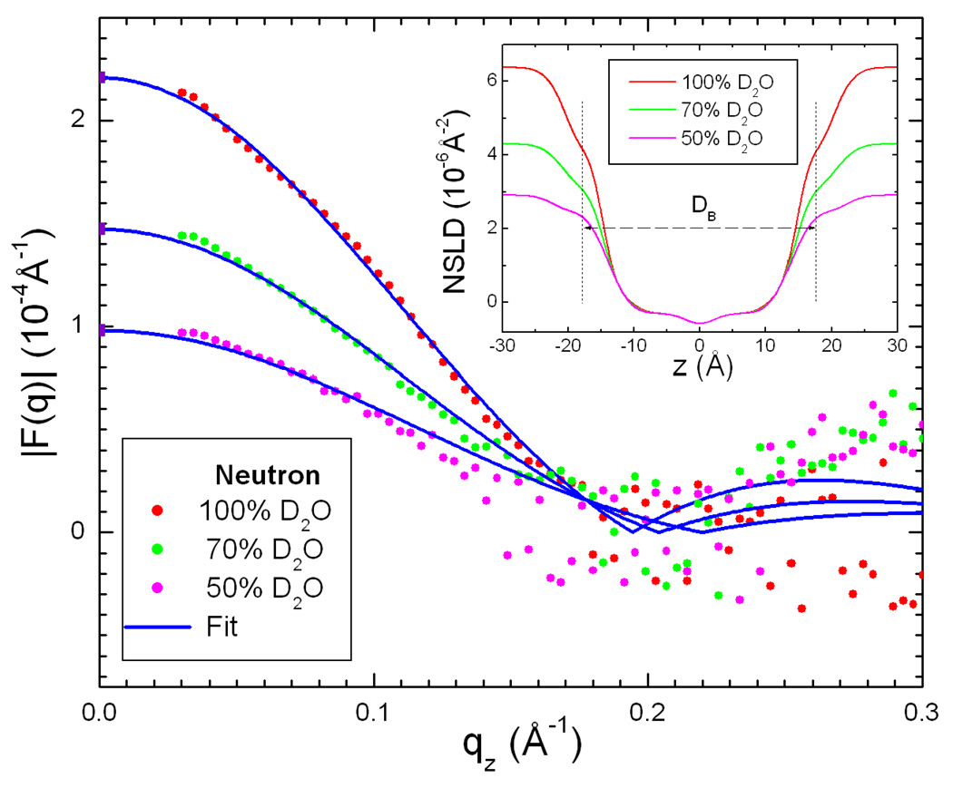
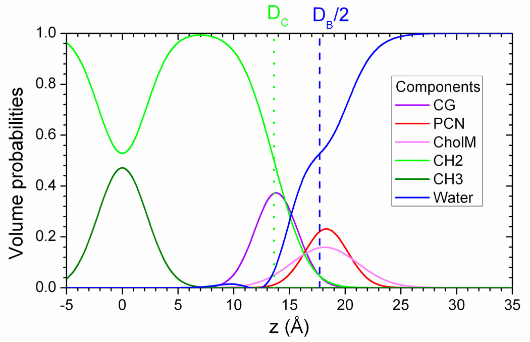
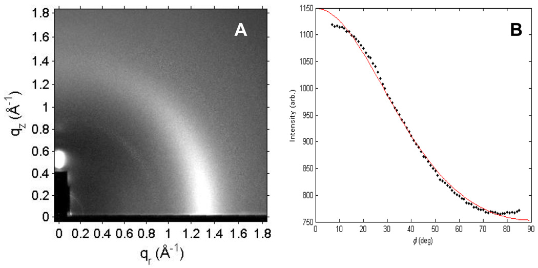
Diphytanoylphosphatidylcholine (DPhyPC) is a branched chain lipid often used for model membrane studies, including peptide/lipid interactions, ion channels and lipid rafts. This work reports results of volume measurements, water permeability measurements P(f), X-ray scattering from oriented samples, and X-ray and neutron scattering from unilamellar vesicles at T=30 degrees C. We measured the volume/lipid V(L)=1426+/-1A(3). The area/lipid was found to be 80.5+/-1.5A(2) when both X-ray and neutron data were combined with the SDP model analysis (Kucerka, N., Nagle, J.F., Sachs, J.N., Feller, S.E., Pencer, J., Jackson, A., Katsaras, J., 2008. Lipid bilayer structure determined by the simultaneous analysis of neutron and X-ray scattering data. Biophys. J. 95, 2356-2367); this is substantially larger than the area of DOPC which has the largest area of the common linear chain lipids. P(f) was measured to be (7.0+/-1.0)x10(-3)cm/s; this is considerably smaller than predicted by the recently proposed 3-slab model (Nagle, J.F., Mathai, J.C., Zeidel, M.L., Tristram-Nagle, S., 2008. Theory of passive permeability through lipid bilayers. J. Gen. Physiol. 131, 77-85). This disagreement can be understood if there is a diminished diffusion coefficient in the hydrocarbon core of DPhyPC and that is supported by previous molecular dynamics simulations (Shinoda, W., Mikami, M., Baba, T., Hato, M., 2004. Molecular dynamics study on the effects of chain branching on the physical properties of lipid bilayers. 2. Permeability. J. Phys. Chem. B 108, 9346-9356). While the DPhyPC head-head thickness (D(HH)=36.4A), and Hamaker parameter (H=4.5x10(-21)J) were similar to the linear chain lipid DOPC, the bending modulus (K(C)=5.2+/-0.5x10(-21)J) was 30% smaller. Our results suggest that, from the biophysical perspective, DPhyPC belongs to a different family of lipids than phosphatidylcholines that have linear chain hydrocarbon chains.


Figures

Figure 1
Figure 1
Chemical structure of DPhyPC (Avanti Polar Lipids image).
Figure 2
Figure 2
Volume/lipid VL vs. molar concentration × of DPhyPC in DLPC determined by neutral buoyancy in D2O/H2O mixtures (red triangles and bottom axis). VL versus weight fraction of DPhyPC in water determined by densimetry (black circles and top axis).
Figure 3
Figure 3
CCD grayscale image of fluid phase LAXS from DPhyPC at 30 °C with higher intensity shown by white pixels and lower intensity by gray pixels. The beam and first lamellar order of the repeat spacing D = 62.5 Å are visible through a semi-transparent molybdenum beam stop that fills the lower left corner.
Figure 4
Figure 4
Bending modulus KC vs. D-spacing for DPhyPC at 30 °C for three samples. The horizontal line shows our estimated value from samples 1 and 3.
Figure 5
Figure 5
Compression modulus B vs. D-spacing for three samples of DPhyPC.
Figure 6
Figure 6
Absolute x-ray form factors (Fourier transforms of the electron density) for DPhyPC at 30 °C. Data from unilamellar vesicles (ULV) and oriented multilayers (ORI) are compared to the SDP model (line). Signs for the phases are indicated for each lobe. Negative values of |F(qz)| indicate statistical fluctuations where scattering intensity is weak, as elaborated previously (Kučerka et al., 2005). The inset shows the electron density profile from the SDP model.
Figure 7
Figure 7
Absolute neutron form factors (Fourier transforms of the scattering length density profile) for ULV of DPhyPC at 30 °C in three concentrations of D2O are compared to the SDP model (lines) with signs of the phases indicated for each lobe. The inset shows the neutron scattering length density profiles.
Figure 8
Figure 8
Volume probabilites for the components of the SDP model: CG (carbonyl+glycerol); PCN (phosphate + 2CH2+N); CholM ((CH3)3 on choline); CH2 (chain methylenes); CH3 (chain terminal methyls); and water (Kučerka et al., 2008). The probabilities are symmetric about the bilayer center at z=0. The dotted green line is located at DC, the Gibbs’ dividing surface for the hydrocarbon region. The dashed blue line is located at DB/2, the Gibbs dividing surface for water.
Figure 9
Figure 9
A. Fluid phase WAXS scattering for DPhyPC at 30 °C (D = 56 Å). B. Intensity vs. φ for the WAXS scattering shown in A. Intensity was integrated in a radial swath over the q range of 0.8 to 1.8 Å−1. The red line is the fit that determines Sxray.
Figure 10
Figure 10
Water permeability Pf at 30 °C of DPhyPC bilayers vs. area/lipid A compared to three linear chain lipids. The open squares show the areas from the older H2 analysis and the solid circles show the areas from the SDP analysis.

References

    1. Bakht O, Pathak P, London E. Effect of the structure of lipids favoring disordered domain formation on the stability of cholesterol-containing ordered domains (lipid rafts): Identification of multiple raft-stabilization mechanisms. Biophys J. 2007;93:4307–4318. - PMC - PubMed
    1. Greenwood AI, Tristram-Nagle S, Nagle JF. Partial molecular volumes of lipids and cholesterol. Chem Phys Lipids. 2006;143:1–10. - PMC - PubMed
    1. Guler SD, Ghosh DD, Pan JJ, Mathai JC, Zeidel ML, Nagle JF, Tristram-Nagle S. Effects of ether vs. ester linkage on lipid bilayer structure and water permeability. Chem Phys Lipids. 2009;160:33–44. - PMC - PubMed
    1. He K, Ludtke SJ, Heller WT, Huang HW. Mechanism of alamethicin insertion into lipid bilayers. Biophys J. 1996;71:2669–2679. - PMC - PubMed
    1. Heller WT, Waring AJ, Lehrer RI, Harroun TA, Weiss TM, Yang L, Huang HW. Membrane thinning effect of the beta-sheet antimicrobial protegrin. Biochemistry-Us. 2000;39:139–145. - PubMed

Publication types

LinkOut - more resources